Epithelioid hemangioendothelioma

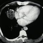
Pulmonary
epithelioid hemangioendothelioma: a case report. Computed tomography scan showing tissular mass with peripheral calcification in the right lung.

Unicentric
epithelioid hemangioendothelioma of the calcaneus: a case report and review of literature. Pre-operative radiograph of large lytic lesion in the calcaneus

Unicentric
epithelioid hemangioendothelioma of the calcaneus: a case report and review of literature. Preoperative sagittal T1 image of the calcaneus showing solid lesion with fairly homogenous signal

Unicentric
epithelioid hemangioendothelioma of the calcaneus: a case report and review of literature. Preoperative sagittal T2 image with some heterogeneity within the lesion surrounding bony edema, sclerotic border around the lesion, but with concern for cortical breakthrough in the subtalar joint

Epithelioid
hemangioendothelioma of the thyroid: a case report. Characteristics of the thyroid nodule. Computed tomography scan showing the right thyroid nodule with calcification (arrow) (a). The nodule is enhanced and shows no signs of extrathyroidal invasion (arrow) (b). Ultrasonogram showing the right thyroid nodule (21 mm in diameter) with calcification (arrow) (c). Macroscopically, a well-circumscribed mass in the right lobe of the thyroid is observed (d)

Hepatic
epithelioid hemangioendothelioma • Hepatic epithelioid hemangioendothelioma - Ganzer Fall bei Radiopaedia

Epithelioid
hemangioendothelioma • Epithelioid hemangioendothelioma - Ganzer Fall bei Radiopaedia

Hepatic
epithelioid hemangioendothelioma • Hepatic and pulmonary epithelioid hemangioendothelioma - Ganzer Fall bei Radiopaedia
Epithelioid hemangioendothelioma (EHE) is a rare relatively low grade vascular tumor. It occurs around medium to large venous structures.
Pathology
It consists of rounded or slightly spindle-shaped eosinophilic endothelial (epitheloid) cells with rounded nuclei and prominent cytoplasmic vacuolisation.
Markers
- factor VII-associated antigen
Location
It can occur in a number of sites
- liver: hepatic epithelioid hemangioendothelioma
- lung: pulmonary epithelioid hemangioendothelioma
- limbs: skin and soft tissues
Radiographic features
Please refer to individual location-specific entities for specific features.
History and etymology
It was initially described by SW Weiss and FM Enzinger in 1982 .
Siehe auch:
- Hämangioendotheliom
- epitheloides Hämangioendotheliom der Leber
- pulmonary epithelioid haemangioendothelioma
- Malignes epitheloides Hämangioendotheliom
- Leberhämangiom bei Kindern
und weiter:

 Assoziationen und Differentialdiagnosen zu epitheloides Hämangioendotheliom:


