hepatic epithelioid hemangioendothelioma


Hepatic epithelioid hemangioendothelioma (HEHE) is a rare, low to intermediate grade malignant hepatic vascular tumor.
Epidemiology
There may be a greater female incidence (with reported male-to-female ratio, 3:2), with peak incidence thought to be around 30-40 years old.
Pathology
Histologically, the tumors are composed of dendritic and epithelioid cells. Tumor cells with intracytoplasmic lumina, occasionally containing red blood cells, appear as signet ring-like structures . They can be difficult to diagnose on the basis of biopsy results.
Radiographic features
General
They tend to be multiple solid tumor nodules, located in a predominantly peripheral distribution, which coalescence to individual nodules. Lesions adjacent to the capsule often produce hepatic capsular retraction.
Ultrasound
Usually seen as hepatic lesions that are predominantly hypoechoic; however, hepatic lesions can also have mixed echotexture or be predominantly hyperechoic.
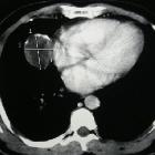
CT
Typically seen as multiple hypoattenuating lesions in both hepatic lobes that coalesce to form larger confluent hypoattenuating regions in a peripheral or subcapsular distribution, with a halo or target pattern of enhancement in larger lesions. Subcapsular lesions often present with capsular retraction. Hepatic or portal veins or their branches may taper and terminate at or just within the edge of these lesions (lollipop sign).
Calcifications are uncommon, but do occur on occasion.
MRI liver
- T1: hypointense lesions relative to normal liver parenchyma on unenhanced T1-weighted images
- T2: heterogeneously increased signal intensity.
- C+ (Gd): some lesions demonstrate either a peripheral halo or a target-type enhancement pattern after administration of a gadolinium-based contrast agent, with an occasional thin peripheral hypointense rim.
Ferumoxide-enhanced T2-weighted images may help physicians distinguish tumor margins
Treatment and prognosis
The clinical course of these lesions can be variable with histologic analysis being of little value in predicting the clinical outcome . The overall prognosis is much more favorable than other hepatic malignancies .
Radical surgical resection or orthoptic liver transplantation are considered the treatments of choice . Due to the often multifocal nature of the tumor, transplantation may be the optimal treatment. Metastatic lesions have been reported in ~30% of patients at presentation and occur most commonly in the lungs . Other less common sites include the abdominal lymph nodes, omentum, mesentery, and peritoneum.
See also
Siehe auch:
- epitheloides Hämangioendotheliom
- Hämangioendotheliom
- hepatic capsular retraction
- seltene Lebertumoren
- Leberhämangiom bei Kindern
und weiter:


 Assoziationen und Differentialdiagnosen zu epitheloides Hämangioendotheliom der Leber: