extrartikuläre Ganglionzyste
MRI
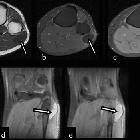
characteristics of cysts and “cyst-like” lesions in and around the knee: what the radiologist needs to know. Common peroneal nerve sheath ganglion cyst. The axial (a) T1-weighted, (b) contrast-enhanced fat saturated T1-weighted, (c) fat saturated proton density weighted images and two sequential coronal (d, e) fat saturated proton density weighted images show a cystic lesion (arrows) at the lateral aspect of the fibula head extending caudally
MRI
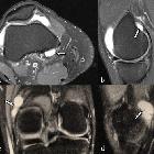
characteristics of cysts and “cyst-like” lesions in and around the knee: what the radiologist needs to know. Extra-articular ganglion cysts. The axial (a) and sagittal (b) fat saturated proton density weighted images demonstrate a unilocular cystic fluid collection consistent with an extra-articular ganglion cyst (arrows). The coronal (c) and sagittal (d) fat saturated proton density weighted images show a multilocular extra-articular ganglion cyst (arrows)
extrartikuläre Ganglionzyste
Siehe auch:


 Assoziationen und Differentialdiagnosen zu extrartikuläre Ganglionzyste:
