zystische Läsionen des Knies

Cyst-like
lesions around the knee • Hoffa's fat pad ganglion cyst - Ganzer Fall bei Radiopaedia

MRI
characteristics of cysts and “cyst-like” lesions in and around the knee: what the radiologist needs to know. Hoffa’s fat pad ganglion cysts: the sagittal (a) and axial (b) fat saturated proton density weighted images in two different patients demonstrate a multiloculated septated cystic lesion (asterisks) within the Hoffa’s fat pad. Ganglion cyst of the suprapatellar bursa: the sagittal (a) and axial (b) fat saturated T2-weighted images show a lobulated cystic lesion (arrows) in the suprapatellar bursa

MRI
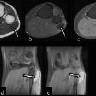
characteristics of cysts and “cyst-like” lesions in and around the knee: what the radiologist needs to know. Common peroneal nerve sheath ganglion cyst. The axial (a) T1-weighted, (b) contrast-enhanced fat saturated T1-weighted, (c) fat saturated proton density weighted images and two sequential coronal (d, e) fat saturated proton density weighted images show a cystic lesion (arrows) at the lateral aspect of the fibula head extending caudally

MRI
characteristics of cysts and “cyst-like” lesions in and around the knee: what the radiologist needs to know. Ganglion cyst of the proximal tibiofibular joint. The coronal fat saturated proton density weighted image shows a ganglion cyst (white arrow) emerging with a short neck (black arrow) from the proximal tibiofibular joint (T tibia, F fibula)

MRI
characteristics of cysts and “cyst-like” lesions in and around the knee: what the radiologist needs to know. Parameniscal cyst. Three sequential sagittal (a–c) fat saturated proton density weighted images demonstrating a lobulated cystic fluid collection (white arrow) in contact with the medial meniscus and arising with a short neck (black arrows) from a horizontal meniscal tear

Cyst-like
lesions around the knee • Giant popliteal artery aneurysm - Ganzer Fall bei Radiopaedia

Cyst-like
lesions around the knee • Parameniscal cyst and lateral meniscal tear - Ganzer Fall bei Radiopaedia

Cyst-like
lesions around the knee • Intra-articular ganglion cyst - knee - Ganzer Fall bei Radiopaedia

Anterior
cruciate ligament ganglion cyst • Anterior cruciate ligament ganglion cyst (intercondylar notch cyst) - Ganzer Fall bei Radiopaedia

Cyst-like
lesions around the knee • Pes anserinus myotendinous injury - Ganzer Fall bei Radiopaedia

MRI
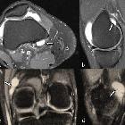
characteristics of cysts and “cyst-like” lesions in and around the knee: what the radiologist needs to know. Synovial sarcoma. The sagittal (a) and axial (b) fat saturated proton density weighted images show a multilocular “cyst-like” lesion near the knee joint (white arrows). The axial (c) T1-weighted and the axial (d) fat saturated contrast-enhanced T1-weighted images aided in the differential diagnosis by showing the solid nature of the tumour. The lesion demonstrated intense contrast enhancement except for a peripherally located necrotic part (grey arrows)

MRI
characteristics of cysts and “cyst-like” lesions in and around the knee: what the radiologist needs to know. ACL ganglion cysts. Two sequential sagittal fat saturated proton density weighted images in two different patients (upper and lower row) demonstrating a cystic lesion (black arrow) at the upper segment of the ACL with part of the lesion (white arrow) dispersing into the ACL fibres

MRI
characteristics of cysts and “cyst-like” lesions in and around the knee: what the radiologist needs to know. PCL ganglion cyst. The sagittal (a) and axial (b) fat saturated proton density weighted images show a multiloculated septated cyst (arrowheads) located in contact with the PCL (white arrows) and along its dorsal surface. Note a small insertional tibial cyst (black arrow)

MRI
characteristics of cysts and “cyst-like” lesions in and around the knee: what the radiologist needs to know. Extra-articular ganglion cysts. The axial (a) and sagittal (b) fat saturated proton density weighted images demonstrate a unilocular cystic fluid collection consistent with an extra-articular ganglion cyst (arrows). The coronal (c) and sagittal (d) fat saturated proton density weighted images show a multilocular extra-articular ganglion cyst (arrows)

MRI
characteristics of cysts and “cyst-like” lesions in and around the knee: what the radiologist needs to know. Intraosseous ganglion cyst. The axial (a) and coronal (b) fat saturated proton density weighted images demonstrate a solitary, unilocular cystic lesion (arrows). Note the sclerotic rim (arrowheads) and mild reactive bone oedema (asterisk)

MRI
characteristics of cysts and “cyst-like” lesions in and around the knee: what the radiologist needs to know. Intrameniscal cysts. The coronal (a) fat saturated proton density weighted image shows a small cystic fluid collection inside the anterior horn of the lateral meniscus (white arrow) communicating with a horizontal meniscal tear (black arrow). The sagittal (b), (c) fat saturated proton density weighted images in two different patients show intrameniscal cysts of the anterior horn of the lateral meniscus (in b) and of the posterior horn of the medial meniscus (in c)

MRI
characteristics of cysts and “cyst-like” lesions in and around the knee: what the radiologist needs to know. Medial parameniscal cyst: axial (a) and coronal (b) fat saturated proton density weighted image demonstrating a parameniscal cyst (asterisk) anterior to the MCL (white arrow) arising from a complex tear of the body of medial meniscus (black arrow). Anterolateral parameniscal cyst: the sagittal (c) fat saturated proton density weighted image shows a parameniscal cyst (small white arrow) extending anteriorly within the Hoffa’s fat pad and continuity with a horizontal tear of the body of the lateral meniscus is noted (black arrow). Lateral parameniscal cyst: axial (d) and two sequential coronal (e and f) fat saturated proton density weighted images demonstrate a parameniscal cyst (asterisk) deep to iliotibial tract (thick white arrows) communicating with a lateral meniscal horizontal tear (black arrows)

MRI
characteristics of cysts and “cyst-like” lesions in and around the knee: what the radiologist needs to know. Prepatellar bursitis. The axial (a), sagittal (b) fat saturated proton density weighted and the sagittal (c) T1-weighted images demonstrate a distended prepatellar bursa (asterisk)

MRI
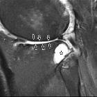
characteristics of cysts and “cyst-like” lesions in and around the knee: what the radiologist needs to know. Subarticular cyst (geode). The sagittal fat saturated proton density weighted image shows a cystic lesion (asterisk) in the subarticular surface of the lateral tibial condyle at the proximal tibiofibular joint. Note chondral defects of the tibial-femoral articular surfaces (black arrows)

MRI
characteristics of cysts and “cyst-like” lesions in and around the knee: what the radiologist needs to know. Semimembranosus insertional cyst. The coronal (a), sagittal (b) and axial (c) fat saturated proton density weighted images show an intraosseous cyst (white arrows) located at the semimembranosus insertion. Semimembranosus tendon is shown with a black arrow. Findings of ACL reconstruction are also visible

MRI
characteristics of cysts and “cyst-like” lesions in and around the knee: what the radiologist needs to know. Popliteal cysts. The axial (a) and sagittal (b) fat saturated proton density weighted images show a large multiseptated popliteal cyst (asterisks) emerging between the medial gastrocnemius tendon (black arrow) and the semimembranosus tendon (white arrow) and abutting the medial gastrocnemial muscle belly (GN). The axial (c) fat saturated proton density weighted image shows a ruptured popliteal cyst (arrowheads). The axial (d) fat saturated proton density weighted image demonstrates a Baker’s cyst (white arrow) with a single loose osteocartilaginous body inside the cyst (black arrow)

MRI
characteristics of cysts and “cyst-like” lesions in and around the knee: what the radiologist needs to know. Suprapatellar bursa. The sagittal (a) and coronal (b) fat saturated proton density weighted images shows the suprapatellar bursa (white arrow) with a partially perforated suprapatellar plica (black arrow)

MRI
characteristics of cysts and “cyst-like” lesions in and around the knee: what the radiologist needs to know. Deep infrapatellar bursitis. The sagittal fat saturated proton density weighted image demonstrates a small fluid collection in the deep infrapatellar bursa (arrow), between the distal patellar tendon and the tibia

MRI
characteristics of cysts and “cyst-like” lesions in and around the knee: what the radiologist needs to know. Pes anserinus bursitis. The axial (a) and coronal (b, c) fat saturated proton density weighted images show a cystic fluid collection (noted with asterisk in a and arrows in b, c) located between the medial aspect of the tibia and the pes anserinus tendons: sartorius muscle (grey arrow), gracilis tendon (white arrow) and semitendinosus tendon (black arrow). T tibia

MRI
characteristics of cysts and “cyst-like” lesions in and around the knee: what the radiologist needs to know. MCL bursitis. The coronal fat saturated proton density weighted image demonstrates a small fluid collection between the deep MCL (meniscofemoral ligament: grey arrow, meniscotibial ligament: black arrow) and the superficial portion (white arrow) of the MCL

MRI
characteristics of cysts and “cyst-like” lesions in and around the knee: what the radiologist needs to know. Semimembranosus-medial collateral ligament bursitis. The axial (a–d) fat saturated proton density weighted images show a fluid distended semimembranosus–MCL bursa (white arrow). In contact with the semimembranosus-MCL bursa a small Baker cyst is demonstrated (black arrow). In image (b) the lesion takes the typical configuration of a distended semimembranosus-MCL bursa, that of an inverted U

MRI
characteristics of cysts and “cyst-like” lesions in and around the knee: what the radiologist needs to know. LCL bursitis. The axial (a) and coronal (b) fat saturated proton density weighted images show a fluid collection in the dilated LCL bursa (asterisk). The white arrow points to LCL and the black arrow points to biceps femoris tendon

MRI
characteristics of cysts and “cyst-like” lesions in and around the knee: what the radiologist needs to know. Knee recesses. The sagittal (a) fat saturated proton density weighted image shows fluid in the subpopliteal-subgastrocnemius (white arrow) and posterior femoral recess (black arrow). Findings of ACL reconstruction are also visible (in dotted line). The axial (b) fat saturated proton density weighted image shows a fluid distended posterior capsular recess which is located posteriorly to the PCL. The axial (c) fat saturated proton density weighted image shows fluid in the subpopliteal recess (white arrow). A Baker cyst (with asterisk) and geodes (black arrows) at both tibial condyles are also shown. The axial (d) fat saturated proton density weighted image shows the central recesses (white arrows) with fluid extending medially and laterally deep to the patellar retinacula

MRI
characteristics of cysts and “cyst-like” lesions in and around the knee: what the radiologist needs to know. Knee recesses. The sagittal (a) fat saturated proton density weighted image shows fluid in the suprahoffatic recess (white arrow), in the infrahoffatic recess (black arrow) and in the suprapatellar pouch (grey arrow). A small fluid intensity lesion in the distal femoral metaphysis (in dotted line) represents a small enchondroma. The coronal (b) fat saturated proton density weighted image shows the parameniscal recesses (white arrows) with fluid above and below the lateral meniscal margins at the level of the body

MRI
characteristics of cysts and “cyst-like” lesions in and around the knee: what the radiologist needs to know. Popliteal artery aneurysm: the axial (a) T1-weighted and the sagittal (b) T2-weighted GRE images show a popliteal aneurysm with thrombus and rim-like calcifications. The blood flow creates an artefact (grey arrows) that helps in the diagnosis. Popliteal vein’s varices: the axial (c) and coronal (d, e) fat saturated proton density weighted images demonstrate lobulated cystic lesions (black arrows) in continuity with the popliteal vein consistent with popliteal vein varices. White small arrow in (c) points to the dilated great saphenous vein

MRI
characteristics of cysts and “cyst-like” lesions in and around the knee: what the radiologist needs to know. Haematoma. The sagittal (a) T1-weighted and the sagittal (b) fat saturated proton density weighted images show a fluid collection in front of the patellar tendon with high signal intensity in T1-weighted image consistent with a haematoma

MRI
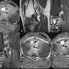
characteristics of cysts and “cyst-like” lesions in and around the knee: what the radiologist needs to know. Brodie’s abscess and lymph nodes in a patient with ACL reconstruction. (a) Axial and (b) sagittal fat saturated proton density weighted, (c) sagittal T1-weighted, (d) sagittal fat saturated contrast-enhanced T1-weighted and (e, f) sequential axial fat saturated contrast-enhanced T1-weighted images. A small cystic lesion (white arrow) at the anterior aspect of the tibia in contact with the ACL graft (black arrow) with marked peripheral enhancement (grey arrows) and a central non-enhancing portion is depicted. The abscess was draining via a sinus tract along the ACL graft to the anterior surface of the tibia. Three small cystic-like lesions (grey arrowheads) in the popliteal fossa, represent enlarged lymph nodes

Cyst-like
lesions around the knee • Hoffa fat pad ganglion cyst - Ganzer Fall bei Radiopaedia
There is broad differential for cyst-like lesions around the knee.
Differential diagnosis
Cysts
- synovial cyst
- ganglion cyst
- intra-articular ganglion cyst
- extra-articular ganglion cyst
- periosteal ganglion cyst
- nerve sheath ganglion cyst
- common peroneal nerve sheath ganglion cyst
- meniscal cyst/parameniscal cyst
- intraosseous cyst
- infective cyst - abscess
Cyst-like lesions
- distended knee bursae
- suprapatellar bursa/suprapatellar bursitis
- prepatellar bursa/prepatellar bursitis
- superficial infrapatellar bursa/superficial infrapatellar bursitis
- deep infrapatellar bursa/deep infrapatellar bursitis
- anserine bursa/pes anserinus bursitis
- iliotibial bursa/iliotibial bursitis
- semimembranosus-tibial collateral ligament (SMTCL) bursa/semimembranosus-tibial collateral ligament (SMTCL) bursitis
- medial collateral ligament bursa/medial collateral ligament bursitis
- fibular collateral ligament-biceps femoris bursa/fibular collateral ligament)-biceps femoris bursitis
- cystic hematoma/post-traumatic cyst
- normal recesses
- posterior femoral recess (subgastrocnemius recess)
- posterior capsular recess
- subpopliteal recess
- suprahoffatic recess
- infrahoffatic recess
- anterior tibial recess
- central synovial recess
- parameniscal recess
- popliteal arterial aneurysm - pseudoaneurysm
- popliteal venous varix
- solid lesions with cyst-like components
- cystic degeneration or myxoid stroma in tumors such as
- synovial sarcoma
- dedifferentiated sarcoma
- myxoid liposarcoma
- metastases
- benign synovial hemangioma
- cystic degeneration or myxoid stroma in tumors such as
Siehe auch:
- Baker-Zyste
- Bursitis am medialen Kollateralband des Knies
- Ganglion des hinteren Kreuzbandes
- Zystische Adventitiadegeneration
- Aneurysma der Arteria poplitea
- Ganglion des vorderen Kreuzbandes
- Knie Ganglion
- Bursitis Knie
- Meniskuszyste
- Ganglion des Hoffa-Fettkörper
- Bursitis am lateralen Kollateralband des Knies
- Bursen des Kniegelenks
- Ganglion des proximalen tibiofibularen Gelenks
- extrartikuläre Ganglionzyste

 Assoziationen und Differentialdiagnosen zu zystische Läsionen des Knies:


