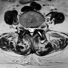
synovial cyst

Facettengelenksarthrose
mit Facettengelenks-Zyste von rechts nach intraspinal mit hochgradiger spinaler Enge.

Intraspinale,
epidurale, eingeblutete Synovialzyste Facettengelenk mit akuter Klinik mit Schmerz und Kaudasymptomatik. Oben sagittal T1 nativ (hell !), T2, STIR, unten T2 axial , T1 KM FS axial und sagittal.

Facettengelenksarthrose
mit Facettengelenks-Zyste nach dorsal. Es findet sich vermehrt Flüssigkeit im Gelenkspalt, links mehr als rechts. Die Zyste schließt sich dorsal links an.

Pseudotumoural
soft tissue lesions of the foot and ankle: a pictorial review. Synovial cyst. A 34-year-old female patient with lateral foot pain and soft tissue mass posterior to the lateral malleolus. Ultrasound examination in the paracoronal (a) and coronal plane (b) demonstrates a well-circumscribed, bilobular, mostly anechogenic lesion (asterisk) with posterior acoustic enhancement, correlating with a cystic nature. Some echogenic debris is present in the dependent part of the lesion. The lesion causes displacement of the sural nerve (arrowheads), causing lateral foot pain

Pseudotumoural
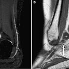
soft tissue lesions of the foot and ankle: a pictorial review. Synovial cyst. Same patient as in Fig. 3. Coronal fat-suppressed T2-WI (a) and sagittal T1-WI (b): well-circumscribed, bilobar lesion (asterisk) with high signal intensity (SI) on T2-WI and low SI on T1-WI. The smaller “lobe” (long arrow) extends caudally towards the posterior facet of the subtalar joint. The cyst causes displacement of the small saphenous vein (short arrow)

Intraspinale
Synovialiszyste, die in Kombination mit anderen degenerativen Frakturen zu einer hochgradigen Spinalkanalstenose führt. Links STIR sagittal, Mitte STIR koronar, rechts T2 axial.

Pseudotumoural
soft tissue lesions of the hand and wrist: a pictorial review. Small cyst (maximum longitudinal size between calliper measurements) at the dorsal aspect of the distal interphalangeal (DIP) joint in a patient with osteoarthritis. Note a small connecting stalk to the adjacent DIP joint (arrow). Longitudinal ultrasound
Synovial cysts are para-articular fluid-filled sacs or pouch-like structures containing synovial fluid and lined by synovial membrane. They can occur around virtually every synovial joint in the body and also around tendon sheaths and bursae. Communication with the adjacent joint may or may not occur.
Siehe auch:
- Bursa iliopectinea
- Ganglion (Überbein)
- Bursitis iliopectinea
- synoviale Zyste Hüftgelenk
- Synovialzysten der Facettengelenke
- eingeblutete Synovialzyste
- eingeblutete Synovialzyste der Facettengelenke
- Facettengelenkszyste HWS
und weiter:
- Facettengelenkszyste LWS
- spinale Arachnoidalzyste
- extraskeletal musculoskeletal tumors by compartment
- Gelenktumoren
- anterior hip pain
- extra skeletal musculoskeletal lesions by compartment
- posttraumatic tendon cyst
- post-traumatic long head of the biceps tendon cyst
- pseudotumoröse Weichteilläsionen von Hand und Handgelenk
- sakrale Arachnoidalzyste
- synovial cyst of the cervical spine
- Sehnenzysten

 Assoziationen und Differentialdiagnosen zu Synovialzyste:


