pseudotumoröse Weichteilläsionen von Hand und Handgelenk

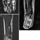
Pseudotumoural
soft tissue lesions of the hand and wrist: a pictorial review. Carpal boss. a Plain radiograph (lateral view) showing a bony prominence at the dorsal aspect of the carpometacarpal joint (arrow). b Sagittal fat-suppressed (FS) TSE T2-weighted image (WI). Note bone marrow oedema and subchondral cyst formation at the carpo-metacarpal joint (arrows)

Pseudotumoural
soft tissue lesions of the hand and wrist: a pictorial review. Extensor carpi ulnaris tenosynovitis. Axial FS T2-WI shows fragmentation into multiple tendon fragments of the extensor carpi ulnaris tendon (arrowheads). Note increased fluid and debris within the tendon sheath

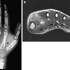
Pseudotumoural
soft tissue lesions of the hand and wrist: a pictorial review. Bizarre parosteal osteochondromatous proliferation (Nora’s lesion). a Plain radiograph of the finger showing a turret exostosis at the dorsal aspect of the proximal phalanx of the second finger (arrowhead). b Longitudinal ultrasound shows the exostosis causing focal contour deformity of the cortical bone (arrowheads) with adjacent hypoechoic cartilage cap (callipers)

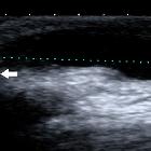
Pseudotumoural
soft tissue lesions of the hand and wrist: a pictorial review. Examples of pseudomasses due to metabolic diseases. a Clinical picture of tophaceous gout. Peri-articular soft-tissue swelling best seen at the proximal interphalangeal joint of the third finger. b Corresponding plain radiograph showing soft-tissue swelling and adjacent periosteal new bone formation (arrow). c Ultrasound of another patient with tophaceous gout at the metacarpophalangeal joint of the thumb, showing a hypoechoic mass with intralesional reflections with retro-acoustic shadowing due to monosodium urate crystal deposition (arrows). d Pseudogout (hydroxyapatite deposition disease) in another patient. Plain radiograph showing linear and amorphous calcifications at the joint capsule of the distal interphalangeal joint of the index (arrows)

Pseudotumoural
soft tissue lesions of the hand and wrist: a pictorial review. Hypothenar hammer syndrome. a Sagittal SE T1-WI shows a hypointense lesion superficial to the flexor tendons (arrowhead) and immediately distal to the hamulus of the hamate (asterisk). b Axial FS SE T1-WI after intravenous administration of gadolinium contrast medium shows focal aneurysmal dilatation of the ulnar artery with central thrombosis (arrow). Note only faint peripheral enhancement. c Doppler ultrasound shows intraluminal thrombosis of the aneurysm of the ulnar artery. d MR angiography shows multiple stenoses at the distal ulnar artery with aneurysmal dilatation with internal clot formation (arrow)
Pseudotumoural
soft tissue lesions of the hand and wrist: a pictorial review. Histopathologically proven rheumatoid nodule in a patient with known rheumatoid arthritis. a Coronal SE T1-WI. Hypointense subcutaneous nodule at the palmar aspect of the distal phalanx of the right digit 3 (arrowheads). b Sagittal FS TSE T2-WI. High signal intensity of the lesion (black arrows). There is moderate pressure erosion of the palmar aspect of the distal phalanx. c Sagittal FS SE T1-WI after intravenous injection of gadolinium contrast medium. There is peripheral enhancement of the lesion (black arrowheads). Palmar localisation of a rheumatoid nodule is less frequent than localisation at the dorsal aspect of the hand

Pseudotumoural
soft tissue lesions of the hand and wrist: a pictorial review. Inflammatory tenosynovitis in a patient with known rheumatoid arthritis. a Transverse ultrasound of the extensor tendons of the wrist showing fluid surrounding the extensor tendons 4. b Transverse power Doppler examination shows increased vascularity within the tendon sheath and tendons

Pseudotumoural
soft tissue lesions of the hand and wrist: a pictorial review. Subcutaneous epidermoid cyst at the phalanges (arrowheads). Longitudinal ultrasound showing a well-defined subcutaneous lesion with variable echogenicity (anechoic components and some internal hyperechoic debris)

Pseudotumoural
soft tissue lesions of the hand and wrist: a pictorial review. Subcutaneous epidermoid cyst with involvement of the adjacent bone of the terminal phalanx of the right fifth finger. a Plain radiograph showing a well-defined osteolytic defect at the radial side of the distal phalanx. Note cortical destruction of the radial cortex. b Coronal SE T1-WI. The lesion is of intermediate signal intensity (arrows) with some internal areas of relatively high signal. c Coronal TSE T2-WI. High signal intensity of the lesion (arrows). d Coronal FS SE T1-WI after intravenous injection of gadolinium contrast medium. There is only subtle peripheral enhancement (arrows) of the lesion

Pseudotumoural
soft tissue lesions of the hand and wrist: a pictorial review. De Quervain tenosynovitis. a Plain radiograph showing a non-specific soft tissue swelling at the radial styloid process (arrow). b Transverse ultrasound showing fluid surrounding the extensor pollicis brevis and abductor pollicis longus. c Longitudinal power Doppler examination shows increased vascularity surrounding the tendons

Pseudotumoural
soft tissue lesions of the hand and wrist: a pictorial review. Small dorsal ganglion cyst. a Axial FS T2-WI. Note a well-delineated hyperintense structure at the dorsal aspect of the wrist (arrow). b The coronal FS TSE T2-WI shows a fluid-filled tear at the scapholunate ligament (arrow)

Pseudotumoural
soft tissue lesions of the hand and wrist: a pictorial review. Foreign body (glass). Longitudinal ultrasound shows multiple hyperechoic reflections with accompanying reverberation artefact (arrows) at the dorsal aspect of the extensor tendons of the fourth digit

Pseudotumoural
soft tissue lesions of the hand and wrist: a pictorial review. Foreign body (wood) crossing the metacarpal heads. a Ultrasound shows a central hyperechoic wood fragment surrounded by a hypoechoic inflammatory reaction. b On power Doppler examination, there is increased vascularity at the periphery of the lesion. c Coronal FS T2-WI, showing a hypointense wooden splinter (arrows), with surrounding high signal band, due to inflammatory reaction. d Axial FS T1-WI after intravenous administration of gadolinium contrast medium showing a rim-enhancing abscess (white arrows) adjacent to the wooden splinter (black arrowhead)
Pseudotumoural
soft tissue lesions of the hand and wrist: a pictorial review. Myositis ossificans of the thenar in a 7-year-old boy presenting with pain and swelling at the thumb (active phase). a Plain radiograph. Note calcifications (arrow) in the thenar. b Axial SE T2-WI shows an ill-defined intramuscular mass that is hyperintense with central areas of low signal. Additionally, there is an intralesional area of heterogeneous low signal, corresponding to the calcifications on plain films (arrow)

Pseudotumoural
soft tissue lesions of the hand and wrist: a pictorial review. Small cyst (maximum longitudinal size between calliper measurements) at the dorsal aspect of the distal interphalangeal (DIP) joint in a patient with osteoarthritis. Note a small connecting stalk to the adjacent DIP joint (arrow). Longitudinal ultrasound

Pseudotumoural
soft tissue lesions of the hand and wrist: a pictorial review. Cyst of the tendon sheath. Longitudinal ultrasound showing a well-delineated anechoic structure (arrow) on the course of the flexor tendon of the right fourth finger (asterisks)
Pseudotumoural
soft tissue lesions of the hand and wrist: a pictorial review. Volar ganglion cyst. Longitudinal ultrasound showing a well-defined anechoic structure at the palmar aspect of the carpus. Note a small stalk-like connection to the adjacent radiocarpal joint (arrow)

Pseudotumoural
soft tissue lesions of the hand and wrist: a pictorial review. Volar ganglion cyst. a Coronal FS T2-WI. Note a polylobular hyperintense cystic structure with a small stalk-like connection to the adjacent scaphotrapezial joint (arrow). b Axial FS T2-WI. The cyst is well delineated and is of high signal intensity (arrowheads)

Pseudotumoural
soft tissue lesions of the hand and wrist: a pictorial review. Palmar fibromatosis. a Longitudinal Doppler ultrasound. Hypoechoic nodule adjacent to the flexor tendon of the palm of the hand (third ray). b Axial SE T1-WI in another patient shows a hypointense lesion at the palmar aspect of the thumb (arrow). c Axial FS SE T1-WI after intravenous administration of gadolinium contrast medium shows diffuse enhancement (arrow)
pseudotumoröse Weichteilläsionen von Hand und Handgelenk
Siehe auch:
- Carpal boss
- Synovialzyste
- Ganglion (Überbein)
- Tendovaginitis stenosans de Quervain
- Epidermoidzyste Finger
- Morbus Dupuytren
- Hypothenar-Hammer-Syndrom
- Fremdkörper in der Hand
- Tendovaginitis Musculus extensor carpi ulnaris
- Weichteilverkalkungen der Hand
- Ganglionzysten der Hand und des Handgelenks
- bizarre parosteale osteochondromatöse Proliferation der Hand und der Finger
- Tendovaginitis Handgelenk
- Myositis ossificans der Handmuskeln
- Sehnenzysten
- Sehnenscheidenzyste


 Assoziationen und Differentialdiagnosen zu pseudotumoröse Weichteilläsionen von Hand und Handgelenk:
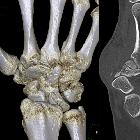


Tendovaginitis
Musculus extensor carpi ulnaris


bizarre
parosteale osteochondromatöse Proliferation der Hand und der Finger