Fehllage Magensonde

Infant with
hematemesis who had an unsuccessful nasogastric tube placementCXR AP shows air outlining the heart in the pericardial space, air outlining the thymus in the mediastinum, and air in the bilateral pleural spaces.The diagnosis was pneumopericardium, pneumomediastinum, and pneumothorax due to esophageal perforation from traumatic nasogastric tube placement. A subsequent esophagram did not demonstrate a leak.


Premature
newborn after replacement of a nasogastric tube. Baseline CXR AP (left) shows the tip of the nasogastric tube to project appropriately over the body of the stomach. CXR AP after nasogastric tube replacement (right) shows the tip of the feeding tube to project over the right upper quadrant of the abdomen.The diagnosis was nasogastric tube malfunction with the tip of the nasogastric tube placed outside the stomach.

Premature
newborn after placement of a nasogastric tube. AXR AP (above left) shows the nasogastric tube to follow a rather straight course into the abdomen. There is increased lucency in the upper abdomen. Subsequent AXR decubitus (above right) shows free air between the abdominal wall and liver. AP view obtained 9 days later immediately after the injection of water soluble contrast through the nasogastric tube (below left) shows some contrast extravasating out of the esophagus into the mediastinum and some contrast entering the stomach. Lateral view obtained 15 minutes later (below right) shows contrast outlining the left pleural space.The diagnosis was nasogastric tube malposition with the nasogastric tube causing esophageal perforation.

Newborn
status post nasogastric tube placementCXR AP shows the tip of the nasogastric tube in the mid esophagus. The tip of the umbilical venous catheter is too high in the right atrium and the tip of the umbilical arterial catheter is too high in the aortic arch.The diagnosis was placement of the nasogastric tube tip in the esophagus.
Teenager with
bilious returns from the nasogastric tubeSupine AXR shows the tip of the feeding tube to be in the gastric body and the tip of the nasogastric tube to be in the third part of the duodenum – the exact opposite of what their normal positions should be.The diagnosis was placement of the nasogastric tube tip post-pyloric in the duodenum.

Fehllage
Magensonde im linken Hauptbronchus. Computertomografie koronarer, sagittal und axial.
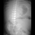
Toddler after
nasogastric tube placement. CXR AP shows the tip of the nasogastric tube to project near the ligament of Treitz.The diagnosis was nasogastric tube malfunction with the tip of the nasogastric tube being transpyloric in position.

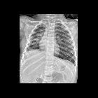
Newborn with
abdominal distension after nasogastric tube placement. CXR AP shows a nasogastric tube curling back upon itself within the esophagus with the tip of the nasogastric tube located about the thoracic inlet.The diagnosis was nasogastric tube malfunction with the nasogastric tube curled in the esophagus.

Asymptomatic
young adult 12 hours after placement of a nasogastric tube during cardiac surgery whose position was not checked after placement. CXR AP shows the tip of the nasogastric tube to be in the left mainstem bronchus.The diagnosis was nasogastric tube malfunction with the tip of the nasogastric tube in the left mainstem bronchus.

Infant after
nasogastric tube placement. CXR AP shows a tracheostomy tube in normal position and a nasogastric tube whose tip is in the right lower lobe.The diagnosis was nasogastric tube malfunction due to placement of the nasogastric tube into the trachea and right mainstem bronchus.
Infant after
nasogastric tube placement. CXR AP shows the tip of the nasogastric tube projects over the second portion of the duodenum.The diagnosis was nasogastric tube malfunction due to transpyloric placement of the tip of the nasogastric tube.


 Assoziationen und Differentialdiagnosen zu Fehllage Magensonde: