Flexor digitorum longus muscle


The flexor digitorum longus (FDL) muscle is located on the tibial side of the leg within the deep posterior compartment of the leg. At its origin it is thin but as it descends, the muscle increases in size.
Summary
- origin: medial side of posterior surface of the tibia
- insertion: plantar surfaces of bases of distal phalanges of the lateral four toes
- action: flexes lateral four toes
- arterial supply: posterior tibial artery
- innervation: tibial nerve
Gross anatomy
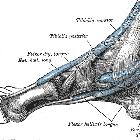
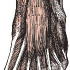
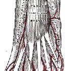
At its origin on the medial distal tibial shaft, it lies medial to the tibialis posterior tendon. The FDL tendon passes through the tarsal tunnel located behind the medial malleolus. It is accompanied by tendons of the tibialis posterior and flexor hallucis longus (FHL) tendons, each occupying a separate synovial-lined tendon sheath . It runs obliquely on the plantar aspect of the foot crossing from medial to lateral. It splits into four smaller tendons each attaching to the base of the distal phalanx of the 2 through 5 toes. As it courses obliquely across the foot, the FDL crosses the deeper placed FHL tendon at a point called the master knot of Henry .
The quadratus plantae muscle attaches onto the lateral aspect of the FDL tendon in the foot.
Variant anatomy
The accessory flexor digitorum longus muscle is an accessory muscle that can occur alongside the flexor digitorum longus muscle. It has a reported prevalence of 6-8% and more commonly occurs unilaterally .
Related pathology
FHL tenosynovitis can occur due to prolonged use requiring extreme or repetitive plantar flexion, e.g. in ballet dancers and marathon runners. Reported points of synovitis include the knot of Henry and the fibro-osseous tarsal tunnel causing pain on the plantar aspect and the posteromedial aspect of the ankle respectively .
The FDL tendon is used in the foot for tendon transfers to correct hammer toe deformities in rheumatoid arthritis .
Siehe auch:
und weiter:

 Assoziationen und Differentialdiagnosen zu Musculus flexor digitorum longus:


