Gallbladder adenoma


Gallbladder adenomas are uncommon gallbladder polyps that, although benign, have a premalignant behavior.
Terminology
As the distinction of adenomas and intracholecystic papillary-tubular neoplasms (ICPN) is not entirely clear, with important overlap between both entities, some authors have proposed that all the adenomas over 1 cm should be grouped under the ICPN terminology . A note is made that the last WHO classification is from 2010, therefore, proceeding these publications.
Epidemiology
Adenomas make 4% to 7% of all gallbladder polyps . They are incidentally found in about 0.5% of the gallbladder specimens .
There is a 2.4:1 female-to-male prevalence ratio .
Associations
Increased prevalence of gallbladder and biliary tract adenomas occurs in :
Pathology
Macroscopic appearance
They are polypoid structures projecting into the gallbladder lumen usually measuring less than 2 cm in size, and showing either a sessile or pedunculated appearance. In about 10% of the cases, adenomas are multiple .
Microscopic appearance
Gallbladder adenomas are classified in :
- tubular adenomas
- the most common
- composed by pyloric-type glands: cuboidal or columnar cells containing vesicular or hyperchromatic nuclei and covered by biliary epithelium
- or by intestinal-type glands: with pseudostratified columnar epithelium covered by biliary epithelium
- papillary adenomas
- papillary structures lined by cuboidal or columnar cells
- tubulopapillary adenomas
- subtype characterized when both the tubular glands and the papillary formations each corresponds to more than 20% of the tumor
Radiographic features
There are no reliable imaging features to distinguish adenomas from gallbladder adenocarcinomas . They might be associated with gallstones or features of chronic cholecystitis.
Ultrasound
Adenomas are usually solitary gallbladder wall lesions that can have a sessile, pedunculated, or polypoid appearance.
- usually hypoechoic with no posterior acoustic shadowing
- variable size, usually between 5 mm to 20 mm
- may have a lobulated or cauliflowerlike contour
- in the pedunculated lesions, the stalk might be difficult to visualize and might require changes in the patient decubitus
- internal vascularity at color Doppler may be demonstrated
- focal gallbladder wall thickening adjacent to the polyp is a worrisome feature concerning for malignancy
- CEUS: enhancement is seen in the arterial phase
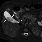
CT
They might be distinguished as small hypodense intraluminal gallbladder lesions that demonstrate enhancement .
Treatment and prognosis
Gallbladder adenomas are usually managed surgically. Please refer to the parental article on gallbladder polyps for guidelines on when followup or surgery should be considered.
Differential diagnosis
- gallbladder adenocarcinoma
- virtually impossible to confiently differentiate on imaging alone
- gallbladder metastasis
Siehe auch:
- Adenomyomatose der Gallenblase
- Polypen der Gallenblase
- tubuläres Adenom der Gallenblase Pylorusdrüsentyp
und weiter:


 Assoziationen und Differentialdiagnosen zu Adenom der Gallenblase:
