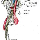
Ganglion oticum


Mandible of
human embryo 95 mm. long. Inner aspect. Nuclei of cartilage stippled.

Mandibular
division of trifacial nerve, seen from the middle line. The small figure is an enlarged view of the otic ganglion. (Testut.)

Plan of the
facial and intermediate nerves and their communication with other nerves.

Diagram of
efferent sympathetic (red) and parasympathetic (blue) nervous system.

Otic ganglion
• Mandibular division of the trigeminal nerve and submandibular and otic ganglia (Gray's illustration) - Ganzer Fall bei Radiopaedia

Otic ganglion
• Autonomic ganglia of the head and neck (Gray's illustrations) - Ganzer Fall bei Radiopaedia
The otic ganglion is one of four parasympathetic ganglia of the head and neck. It receives parasympathetic fibers from the glossopharyngeal nerve.
Gross anatomy
- small and disc shaped
- located in the infratemporal fossa
- it lies immediately below the foramen ovale, medial to the mandibular division of the trigeminal nerve
- supplies secretomotor fibers to the parotid gland.
Roots
- parasympathetic root
- from the inferior salivary nucleus via the glossopharyngeal nerve and branch off the tympanic nerve (Jacobson's nerve) to form the tympanic plexus in the middle ear and then exit as the lesser petrosal nerve. The lesser petrosal nerve then passes from the foramen ovale to the ganglion and then fibers reach the parotid gland as the auriculotemporal nerve.
- sympathetic root
- via the middle meningeal artery, vasomotor fibers from the superior cervical ganglion pass through the ganglion without synapsing and course though the auriculotemporal nerve and supply the parotid gland
- sensory root
- via the auriculotemporal nerve is sensory to the parotid gland.
Branches
Related pathology
Frey syndrome: salivary stimuli will induce perspiration and erythema at the parotid
Siehe auch:
und weiter:

 Assoziationen und Differentialdiagnosen zu Ganglion oticum:

