gastric perforation

Newborn with
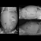
esophageal atresia with abdominal distension after gastrostomy tube placement 1 week agoSupine AXR (left) shows a gastrostomy tube projecting appropriately over the stomach with a triangular lucency superior to the stomach. Left lateral decubitus AXR (above right) again shows the triangular lucency superior to the stomach but does not show air between the abdominal wall and liver. Cross-lateral AXR (below right) shows air between the anterior abdominal wall and liver. The diagnosis was pneumoperitoneum due to gastric perforation after gastrostomy placement. In the operating room the patient was found to have ischemic necrosis of the greater curvature of the stomach.

Newborn with
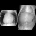
distended abdomen after nasogastric tube placementSupine AXR (left) shows a large amount of air within the abdomen and air outlining both sides of bowel wall (Rigler’s sign) in the right lower quadrant. Supine AXR taken later after pulling back of the nasogastric tube out of the stomach shows visualization of the falciform ligament over the spine (American football sign)The diagnosis was pneumoperitoneum due to gastric perforation during nasogastric tube placement.

Premature
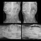
newborn after nasogastric tube placementSupine and left lateral decubitus AXR (left) show a nasogastric tube with its tip deep in the pelvis without evidence of free air. Supine AXR taken after pulling the nasogastric tube back into the stomach (above right) shows increased lucency throughout the central abdomen and left lateral decubitus AXR taken at same time (below right) shows air between the abdominal wall and the liver.The diagnosis was pneumoperitoneum due to gastric perforation during nasogastric tube placement which became visible only after the nasogastric tube was pulled out of the hole it had made in the stomach wall.

Air in the
lesser sac. (a, b) Contrast-enhanced axial CT shows the presence of anomalous gas in the lesser sac, with communication with the gastric lumen (arrow) consistent with a contained perforation.

Air in the
lesser sac. (a, b) Contrast-enhanced axial CT shows the presence of anomalous gas in the lesser sac, with communication with the gastric lumen (arrow) consistent with a contained perforation.

Air in the
lesser sac. (c) Contrast-enhanced coronal CT shows the presence of anomalous gas in the lesser sac (arrow), with communication with the gastric lumen consistent with a contained perforation.

Air in the
lesser sac. (d) Lung window axial CT better depicts the gastric wall discontinuity.
 Assoziationen und Differentialdiagnosen zu Magenperforation:


