gastrointestinal tuberculosis


Tuberculosis
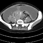
• Retroperitoneal tuberculosis resulting in renal obstruction - Ganzer Fall bei Radiopaedia

Toddler with
abdominal pain who also has a long standing cough. AXR AP shows multiple scattered punctate calcifications just to the right of the L3 and L4 vertebral bodies. A dilated loop of distal small bowel is seen in the left lower quadrant.The diagnosis was gastrointestinal tuberculosis involving multiple abdominal lymph nodes in the right lower quadrant.

Abdominal
tuberculosis • Ileocecal tuberculosis and appendicitis - Ganzer Fall bei Radiopaedia

Abdominal
tuberculosis • Gallbladder tuberculosis - Ganzer Fall bei Radiopaedia

Abdominal
tuberculosis • Disseminated tuberculous infection with miliary disease, lymphadenitis, and colitis - Ganzer Fall bei Radiopaedia

Abdominal
tuberculosis • Cerebral and abdominal tuberculosis (PET-CT) - Ganzer Fall bei Radiopaedia

Abdominal
tuberculosis • Tuberculous lymphadenitis - Ganzer Fall bei Radiopaedia

Tubo-ovarian
abscess • Tuberculous tubo-ovarian abscess and peritonitis - Ganzer Fall bei Radiopaedia

Splenic
granulomatous disease • Splenic and hepatic tuberculous granulomatosis - Ganzer Fall bei Radiopaedia

Abdominal
tuberculosis • Abdominal wall and retroperitoneal tuberculosis - Ganzer Fall bei Radiopaedia

Abdominal
tuberculosis • Tuberculous lymphadenitis - abdomen - Ganzer Fall bei Radiopaedia


Extrapulmonary
tuberculosıs: an old but resurgent problem. A 31-year-old female. Contrast-enhanced CT image demonstrates diffuse-symmetric wall thickening and enhancement of the cecum with surrounding inflammatory changes (arrows)

Extrapulmonary
tuberculosıs: an old but resurgent problem. Transverse CT image without intravenous contrast of a 24-year-old female. Diffuse-symmetric wall thickening of the ileal segment is noted (arrows). Ileal TB

Extrapulmonary
tuberculosıs: an old but resurgent problem. A 40-year-old female. Contrast-enhancement CT scan (a, b) and Doppler ultrasonography (c) demonstrated a large heterogeneous cystic-necrotic mass in the head of the pancreas

Extrapulmonary
tuberculosıs: an old but resurgent problem. A 33-year-old female. Axial post-contrast CT images (a, b) demonstrate omental nodularity, mesenteric fat stranding (arrowheads), and ascites (star). Intraabdominal lymphadenopathies are also evident (arrows) in image b

Extrapulmonary
tuberculosıs: an old but resurgent problem. A 24-year-old male patient. Axial (a) and coronal postcontrast CT (b) images show mesenteric striation (arrowheads) and ascites (star)

Extrapulmonary
tuberculosıs: an old but resurgent problem. Transverse CT image with intravenous contrast of a 29-year-old male. Massive ascites is indicated in the abdomen cavity (stars). The peritoneum"s thin linear contrast enhancement is also noted. Tuberculous peritonitis

Extrapulmonary
tuberculosıs: an old but resurgent problem. A 19-year-old male. Post-contrast CT image shows multiple mesenteric lymphadenopathies with peripheral enhancement


Toddler with
new abdominal pain who also has had a long standing cough. AP image from late in a small bowel follow through exam shows contrast exiting the small bowel and beginning to fill the cecum and ascending colon. The terminal ileum in the right lower quadrant is narrowed with only a thin string of contrast within it. The cecal pole is also somewhat narrowed in appearance.The diagnosis was gastrointestinal tuberculosis involving the terminal ileum.

Abdominal tuberculous can manifest in almost every abdominopelvic organ:
- gastrointestinal tuberculosis
- esophageal tuberculosis
- gastric tuberculosis
- duodenal tuberculosis
- jejunal and ileal tuberculosis
- ileocecal tuberculosis
- colorectal tuberculosis
- tuberculous peritonitis
- tubercular lymphadenopathy
- visceral tuberculosis
- hepatic tuberculosis
- gallbladder tuberculosis
- pancreatic tuberculosis
- splenic tuberculosis
- genitourinary tuberculosis
- renal tuberculosis
- bladder and ureteric tuberculosis
- prostatic tuberculosis
- scrotal tuberculosis (testes, epididymis, seminal vesicles, vas deferens)
- tuberculous pelvic inflammatory disease (female)
Pathology
There are three main pathways for tuberculous infection of the abdomen :
- ingestion of infected milk or sputum initially affects gastrointestinal tract mucosa, followed by the remainder of the bowel wall, regional lymph nodes and peritoneum
- haematogenous spread to the peritoneum, lymph nodes and solid viscera
- direct spread to the peritoneum, e.g. from skeletal tuberculosis via a psoas abscess
Siehe auch:
- Tuberkulose des Peritoneums
- Tuberkulose des Pankreas
- mesenteric panniculitis associated with abdominal tuberculous lymphadenitis
- Tuberkulose des Ösophagus
- intra-abdominal abscess due to Mycobacterium Tuberculosis
- abdominelle tuberkulöse Lymphadenitis
und weiter:
- Fleischner sign (tuberculosis of ileocecal junction)
- granulomatöse Peritonitis
- Peritonitis chronica fibrosa incapsulata
- peritoneal tuberculosis following infliximab therapy
- duodenal tuberculosis
- peritoneal tuberculosis: CT evaluation
- Tuberkulose des Peritoneums bei Kindern
- Tuberkulose Nebenniere
- ileozökale Tuberkulose
- biliäre Tuberkulose
- Stierlin-Zeichen


 Assoziationen und Differentialdiagnosen zu intraabdominelle Tuberkulose: