gastrojejunostomy tube malfunction
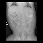
Teenager who
is having emesis during feeding through the jejunostomy port of his gastrojejunostomy tube. AXR taken after injection through the jejunostomy port of a newly placed gastrojejunostomy tube one month ago (above) shows the tip of the jejunostomy tube to be in the proximal jejunum. AXR taken today (below) shows that the tip of the jejunostomy tube has been pulled back into the antrum of the stomach.The diagnosis was gastrojejunostomy tube malfunction due to migration of the tip of the jejunostomy tube back into the stomach.

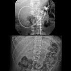
Young adult
with a gastrojejunostomy tube and chronic abdominal pain. Axial (above) CT with contrast of the abdomen shows a round soft tissue mass to the right of the vertebral body that has a target sign appearance and that has a jejunostomy tube coursing in the center of it. Coronal CT (below) shows the soft tissue mass to be long in length and to comprise the second and third parts of the duodenum and the proximal jejunum and to have the jejunostomy tube coursing throughout its length.The diagnosis was gastrojejunostomy tube malfunction due to the formation of a duodenal-jejunal intussuception forming around the tip of the jejunostomy tube.
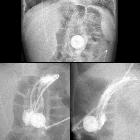
Toddler with
problems feeding through the jejunostomy port of their gastrojejunostomy tube. AXR (above) shows the tip of the jejunostomy tube projects over the body of the stomach. AP (below left) and lateral (below right) images obtained after injecting water soluble contrast through the jejunostomy port of the gastrojejunostomy tube shows contrast outlining the rugae of the stomach. There is no contrast in the duodenum or jejunum.The diagnosis was gastrojejunostomy tube malfunction with the tip of the jejunostomy tube having been pulled back into the stomach.
gastrojejunostomy tube malfunction
Siehe auch:


 Assoziationen und Differentialdiagnosen zu gastrojejunostomy tube malfunction: