Fehlfunktion Ernährungssonde

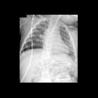
Infant after
feeding tube placementAXR obtained immediately after feeding tube placement (left) shows a feeding tube going down the left mainstem bronchus and then turning up into the lung and increased lucency in the left costophrenic angle presumably due to the feeding tube entering the left pleural space. AXR obtained a minute later after feeding tube repositioning (right) shows the tip of the feeding tube in the antrum of the stomach and a large left pleural air collection with mediastinal shift to the right.The diagnosis was feeding tube malfunction due to placement of the feeding tube through the airway into the lung and pleural space causing a tension pneumothorax.
Toddler after
feeding tube placementAXR shows a feeding tube entering the stomach and then looping back upon itself into the proximal esophagus.The diagnosis was feeding tube malfunction due to placement of the feeding tube tip in the esophagus.
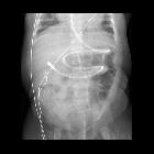
Preschooler
after feeding tube placementAXR shows a feeding tube coursing along the greater curvature of the stomach with the tip looping back upon itself in the body of the stomach. A nasogastric tube is also present with its tip within the body of the stomach.The diagnosis was feeding tube malfunction due to placement of the feeding tube tip in the stomach.

Infant after
feeding tube placementAXR shows a feeding tube coursing through the stomach and then passing through the pylorus with the tip in the second part of the duodenum. A nasogastric tube is also present with its tip within the fundus of the stomach.The diagnosis was feeding tube malfunction due to placement of the feeding tube tip in the second part of the duodenum.
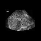
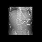
Preschooler
with superior mesenteric artery syndrome who had a feeding tube placed into the third part of the duodenum 1 day ago that is now not working. AXR (not available) showed the tip had migrated into the right upper quadrant of the abdomenTransverse US of the liver shows the echogenic tip of a feeding tube to be in the main portal vein in the center of the image. Note the posterior shadowing from the tip extending inferiorly from it.The diagnosis was feeding tube malfunction due to migration of the feeding tube tip out of the duodenum into the main portal vein. In the operating room the feeding tube was seen to have eroded into the superior mesenteric vein and then into the main portal vein and it was removed without complication.
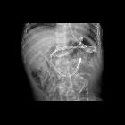
Infant having
difficulties with tube feedings. AXR AP (above) shows a feeding tube that crosses to the right of the spine and then heads inferiorly before turning back to the left of the spine with its tip projecting in the left upper quadrant and this is also demonstrated on the pre-tube injection scout image (below left). Injection of contrast through the tube (below right) showed the tip was in the stomach.The diagnosis was feeding tube malfunction with pulling back of the feeding tube tip out of the duodenum and into the stomach.
Infant whose
feeding tube cannot be flushed and fed through. CXR AP shows a feeding tube with its tip projecting over the proximal jejunum. Incidentally noted is an aneurysmal dilation of the feeding tube just beneath the level of the thoracic inlet. The diagnosis was feeding tube malfunction due to aneurysmal dilation of its proximal portion.

Toddler after
feeding tube placement. CXR AP shows a feeding tube which courses into the stomach and then loops back upon itself and reenters the esophagus and whose tip lies at the level of C6.The diagnosis was feeding tube malfunction with malposition of the feeding tube tip in the cervical esophagus.
Infant who is
having difficulty being fed through their feeding tube. AXR AP shows the tip of the nasogastric tube to be in the body of the stomach. The tip of the feeding tube is transpyloric in position at the duodenal jeujunal junction. However, the feeding tube lumen is kinked in the body of the stomach.The diagnosis was feeding tube malfunction due to a kink in the feeding tube.
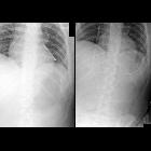
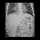
Young adult
who is coughing after feeding tube placement. Initial AXR (left) shows the tip of the feeding tube in the left mainstem bronchus. Subsequent AXR after feeding tube repositioning (right) shows the feeding tube tip to be in the fundus of the stomach.The diagnosis was feeding tube malfunction with the feeding tube tip in the left mainstem bronchus.
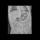
Infant who
cannot be fed through their feeding tube. AXR AP shows the tip of the feeding tube to be at the duodenal-jejunal junction. In the antrum of the stomach the feeding tube is curled back upon itself and kinked at this location.The diagnosis was feeding tube malfunction due to a kink in the feeding tube.
Fehlfunktion Ernährungssonde
Siehe auch:
und weiter:


 Assoziationen und Differentialdiagnosen zu Fehlfunktion Ernährungssonde: