Glass foreign bodies


Glass foreign bodies may be present if they are ingested, inserted, or as a result of an injury. All glass is radiopaque .
Epidemiology
The prevalence of glass foreign bodies in wounds from injury has been recorded at a rate of 1.5% in superficial (subcutaneous) wounds and 7.5% of deeper wounds . They make up ~15% (range 9-24%) of all retained foreign bodies .
Radiographic features
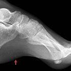
Plain radiograph
Only ~10% of x-rays ordered for investigation of retained foreign bodies are positive (reflecting the low incidence post-injury), but x-rays are excellent at detecting radiopaque foreign bodies with ~85% being detected :
- glass is always radiopaque, independent of lead content or other additives, with the caveat that tiny pieces may be too small to actually be resolved (see below)
- should be visible on plain films if larger than 2 mm
CT
All glass is visible on CT and usually easier to see than on plain radiography . Density varies between 500-1900 HU. Dense fragments as small as 0.01 mm can be detected .
Ultrasound
- can be used to localize foreign bodies further and define the relationship with soft tissue structures and assess for further injuries
- appears hyperechoic with posterior acoustic shadowing and often demonstrates reverberation artifact
- if present for >24 hours may demonstrate a hypoechoic ring
MRI
MRI would clearly not be the first choice investigation for detecting foreign bodies, including glass. Nevertheless, on MRI all forms of glass are seen but on most sequences considerable artifact is present .
Signal characteristics
- T1: low signal
- T2: low signal
- T2*: blooming artifact
- T1C+: linear enhancement if a foreign body granuloma has formed
Siehe auch:
und weiter:


 Assoziationen und Differentialdiagnosen zu Fremdkörper Glas:
