Gleichgewichtsorgan
This image
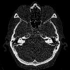
is part of a series which can be scrolled interactively with the mousewheel or mouse dragging. This is done by using Template:Imagestack. The series is found in the category Temporal bone in computertomography case 001. Felsenbein in der Computertomographie. Bilder für scrollbaren Stapel.

desc}} ==
{{Information |description = |Other fields 1 = {{Information field|Name={{Ucfirst:{{Plate}}}}|Value=901}} |date = {{Other date|before|1858}} |source = *{{cite book |year=1918 |author=[[:en:Henry Gray|Henry Gray]] |title=Anatomy of the Human Body }} (See "{{Section header|Book}}" section below) *{{Gray"s Anatomy/link|901}} |author = {{Creator:Henry Vandyke Carter}} |other_versions = |permission = }} == {{Section header|Book}} == {{Gray"s Anatomy}} =={{int:license-header}}== {{PD-scan|PD-old-100-1923}} [[Category:Gray"s Anatomy plates|0901]] [[Category:Anatomical plates and drawings of the


Position of
the right bony labyrinth of the ear in the skull, viewed from above. The temporal bone is considered transparent and the labyrinth drawn in from a corrosion preparation. (Spalteholz.)


Right human
membranous labyrinth, removed from its bony enclosure and viewed from the antero-lateral aspect. (G. Retzius.)

The same from
the postero-medial aspect. 1. Lateral semicircular canal; 1’, its ampulla; 2. Posterior canal; 2’, its ampulla. 3. Superior canal; 3’, its ampulla. 4. Conjoined limb of superior and posterior canals (sinus utriculi superior). 5. Utricle. 5’. Recessus utriculi. 5”. Sinus utriculi posterior. 6. Ductus endolymphaticus. 7. Canalis utriculosaccularis. 8. Nerve to ampulla of superior canal. 9. Nerve to ampulla of lateral canal. 10. Nerve to recessus utriculi (in Fig. 925, the three branches appear conjoined). 10’. Ending of nerve in recessus utriculi. 11. Facial nerve. 12. Lagena cochleæ. 13. Nerve of cochlea within spiral lamina. 14. Basilar membrane. 15. Nerve fibers to macula of saccule. 16. Nerve to ampulla of posterior canal. 17. Saccule. 18. Secondary membrane of tympanum. 19. Canalis reuniens. 20. Vestibular end of ductus cochlearis. 23. Section of the facial and acoustic nerves within internal acoustic meatus (the separation between them is not apparent in the section). (G. Retzius.)

Transverse
section of a human semicircular canal and duct (after Rüdinger).
Gleichgewichtsorgan
Siehe auch:
und weiter:


 Assoziationen und Differentialdiagnosen zu Gleichgewichtsorgan: