Hämangiom der Cauda equina

Capillary
hemangioma of cauda equina: a case report. Plain radiography of lumbar area showing bony erosion and widening of the canal (scalloping) at the level of L3 vertebra (arrow).

Capillary
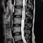
hemangioma of cauda equina: a case report. Sagital magnetic resonance images showing an intradural mass at the level of L3, isointense to spinal cord on T1-weighted (A) and iso-hyperintense on T2-weighted sequences (B). T1-weihted axial image demonstrating the intradural mass pushing the caudal roots to the side of the canal (C).

Capillary
hemangioma of cauda equina: a case report. Post-gadolinium enhancement revealed in sagital (A), coronal (B), and axial (C) magnetic resonance images of this lesion.


 Assoziationen und Differentialdiagnosen zu Hämangiom der Cauda equina: