Histoplasmose

Teenager with
cough, shortness of breath, and chest painCT with contrast of the chest shows bilateral hilar adenopathy.The diagnosis was histoplasmosis.

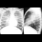
School ager
with chronic cough. CXR PA and lateral shows bilateral hilar lymphadenopathy, right greater than left, and small miliary nodules throughout the lungs.The diagnosis was histoplasmosis.

Thoracic
histoplasmosis • Acute miliary histoplasmosis - Ganzer Fall bei Radiopaedia

Teenager with
hypertension. Coronal CT with contrast of the chest (left) shows a large round soft tissue nodule with a calcified center in the lateral aspect of the left hemithorax. Axial CT (above right) shows calcified mediastinal lymph nodes near the descending aorta and better demonstrates the calcification in the lung nodule (below right).The diagnosis was histoplasmosis.

Histoplasmosis
• Adrenal histoplasmosis - Ganzer Fall bei Radiopaedia

School ager
with 1 week of abdominal pain and shortness of breath. AXR (above left) shows hepatomegaly and an enlarged cardiac silhouette. CXR (above right) shows a water-bottle appearance to the cardiac silhouette and bilateral pleural effusions and bilateral hilar lymphadenopathy. Transverse US of the heart (below) shows a large anechoic fluid collection in the pericardial space.The diagnosis was pericardial effusion due to histoplasmosis.

Thoracic
histoplasmosis • Disseminated histoplasmosis - Ganzer Fall bei Radiopaedia

School ager
with shortness of breath. CXR AP (above left) shows a large cardiac silhouette and an abnormal contour to the right superior mediastinum. Coronal CT with contrast of the chest (above right) shows a huge fluid collection in the pericardial space and a conglomeration of cystic lymph nodes in the right superior mediastinum. Axial CT (below) shows the pericardial fluid collection completely surrounding the heart and left lower lobe atelectasis.The diagnosis was pericardial effusion due to histoplasmosis.

Disseminated
histoplasmosis, a rare cause of abdominal lymphadenopathy. Abdominal ultrasound (a,b) detected a few ovoid-shaped lymphadenopathies (calipers, 3 cm maximal diameter) with thickened hypoechoic cortex and thin central hilum.

Disseminated
histoplasmosis, a rare cause of abdominal lymphadenopathy. Neck ultrasound showed confluent adenopathies with variable echogenicity on the right side (caliper in c), a larger left-sided strongly hypoechoic nodal mass (caliper in d) with nodular margins measuring 36 x 19 mm.

Disseminated
histoplasmosis, a rare cause of abdominal lymphadenopathy. Panoramic coronal image showed bilateral, partically confluent neck adenopathies, multiple mesenterial, retroperitoneal and iliac adenopathies in the abdomen, either homogeneous (arrows) or centrally necrotic (arrowheads).

Disseminated
histoplasmosis, a rare cause of abdominal lymphadenopathy. Axial neck images (f...h) showed several, partially confluent bilateral adenopathies, , either homogeneously enhancing (arrows) or centrally necrotic (arrowheads).
Histoplasmosis is an endemic mycosis caused by Histoplasma capsulatum.
Pulmonary histoplasmosis is the most common manifestation of this infectious disease. Disseminated/extrapulmonary (pericardial, articular) histoplasmosis is often seen in immunosuppressed patients. As such, these are included amongst the infectious etiologies of AIDS-defining illnesses.
See also
Siehe auch:
und weiter:

 Assoziationen und Differentialdiagnosen zu Histoplasmose:
thorakale
oder pulmonale Histoplasmose
