hutch diverticulum


 nicht verwechseln mit: Ureterozele
Hutch diverticula are congenital bladder diverticula, seen at the vesicoureteric junction, in the absence of posterior urethral valves or neurogenic bladder. They are thought to result from a weakness in the detrusor muscle anterolateral to the ureteral orifice.
Epidemiology
They occur almost exclusively in boys with an estimated prevalence of approximately 2% .
Clinical presentation
Urinary tract infection, incontinence, or urinary retention are the most common presentations.
Pathology
Hutch diverticula are commonly seen with vesicoureteric reflux, because the normal oblique insertion of the ureter is disrupted.
Associations
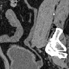
Radiographic features
Fluoroscopy
On voiding cystourethrogram, contrast-filled outpouchings from the urinary bladder arise at the vesicoureteric junction, giving "Mickey Mouse appearance" . Vesicoureteral reflux is often associated.
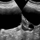
Ultrasound
Round or oval anechoic fluid collections arising from the base of the urinary bladder or around the ureteric orifice .
Treatment and prognosis
Incidentally detected diverticula usually do not require any therapy. If associated with reflux, recurrent infection, or obstruction, they should be resected. However, because of the risk of the development of carcinoma, some surgeons advocate prophylactic surgery in all cases. Hutch diverticula are prone to spontaneous rupture, as well as post-surgical recurrence.
History and etymology
Primary and secondary paraureteric diverticula were first described by American urologist John Hutch (-1972) in 1961 .
Differential diagnosis
- "bladder ears"
- these are transient outpouching normally seen in infants, and represent normal protrusions of urinary bladder into the inguinal rings
- lateral voiding cystourethrogram usually clarifies the diagnosis
See also
Siehe auch:
- Ureterozele
- Harnblasendivertikel
- beidseitiges periureterales Divertikel der Harnblase (Hutch-Divertikel)
und weiter:


 Assoziationen und Differentialdiagnosen zu periureterales Divertikel der Harnblase (Hutch-Divertikel):

