hypovolemic shock

Hyperdense,
hyperperfundierte Nebennieren nach abdomineller Hypoperfusion im Rahmen eines septischen Geschehens. Computertomographie axial in arterieller und portalvenöser Kontrastmittelphase.

CT findings
in a typical hypovolaemic shock. Axial CT acquisition after IV contrast administration depicts the presence of gaseous bubbles in the intrahepatic bile ducts due to aerobilia (arrows).

CT findings
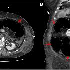
in a typical hypovolaemic shock. (A) Axial CT acquisition and (B) coronal reformation after IV iodinated contrast administration show the presence of luminal gas in the sigmoid, ascending and transverse colon causing bowel distention (arrows).

CT findings
in a typical hypovolaemic shock. Axial CT acquisitions after contrast administration demonstrating a flattening of the inferior vena cava that has a caliber of only 4 mm at the renal arteries level (A) and 3 mm measured 2 cm below them (B).

CT findings
in a typical hypovolaemic shock. Axial non-contrast (A) and coronal (B) reformation after administration of IV contrast demonstrate well-defined peripheral areas of liver parenchyma"s unenhancement at segments II (red), VIII (blue), VIII / V (green) and VI (yellow).

CT findings
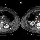
in a typical hypovolaemic shock. Axial (A) and coronal (B) CT reformations after IV iodinated contrast administration reveal an increased enhancement of both adrenal glands after intravenous contrast administration (arrows).

CT findings
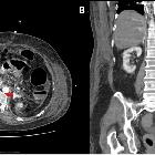
in a typical hypovolaemic shock. Axial CT reformations before (A) and after (B) IV iodinated contrast administration depicting an increased enhancement of renal parenchyma (arrows).

CT findings
in a typical hypovolaemic shock. Coronal reformations in the arterial phase shows retrograde opacification of the hepatic veins and of the vena cava axis (red arrows). Loculated ascites (blue arrow).

Hypovolemic
shock with subtle imaging signs: systemic capillary leak syndrome. Sagittal reformatted image confirmed slit-like retrohepatic and infrahepatic IVC (arrows) with sub-centimeter anteroposterior diameter.

Hypovolemic
shock with subtle imaging signs: systemic capillary leak syndrome. The extremely thin retrohepatic IVC (arrow) was surrounded by subtle hypoattenuating oedematous halo.

Hypovolemic
shock with subtle imaging signs: systemic capillary leak syndrome. Similarly to the slit-like IVC (arrow), the renal veins also appeared collapsed.

 Assoziationen und Differentialdiagnosen zu hypovolämischer Schock:
