Kapillarlecksyndrom

Hypovolemic
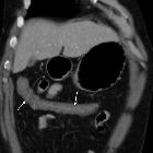
shock with subtle imaging signs: systemic capillary leak syndrome. Most of the collapsed colon showed diffuse, moderate circumferential mural thickening (thin arrows) with mucosal enhancement and mural hypoattenuation suggesting submucosal oedema.

Hypovolemic
shock with subtle imaging signs: systemic capillary leak syndrome. Most of the collapsed colon showed diffuse, moderate circumferential mural thickening (thin arrows) with mucosal enhancement and mural hypoattenuation suggesting submucosal oedema.

Hypovolemic
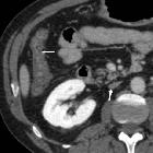
shock with subtle imaging signs: systemic capillary leak syndrome. Diffuse, moderate circumferential mural thickening (thin arrows) with mucosal enhancement and mural hypoattenuation suggesting submucosal oedema. Note marked nephrogram, slit-like inferior vena cava (arrow).

Hypovolemic
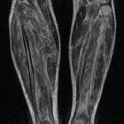
shock with subtle imaging signs: systemic capillary leak syndrome. Coronal (a,b) and axial (c) T2-weighted images showed hyperintense oedematous muscle swelling of both calves, particularly severe on the right side and mostly affecting the gastrocnemius and soleus muscles.

Hypovolemic
shock with subtle imaging signs: systemic capillary leak syndrome. Axial fat-suppressed T2-weighted images (d,e) better showed intramuscular oedematous changes, with associated fluid-like epimysial, perimysial and fascial effusions, causing swelling particularly severe in the right calf.

Hypovolemic
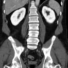
shock with subtle imaging signs: systemic capillary leak syndrome. Portal venous phase images showed marked, bilateral symmetric and homogeneous enhancement (average 320 Hounsfield units compared to 35 HU precontrast attenuation) of the renal parenchyma.
Capillary leak syndrome is a situation characterized by the escape of blood plasma through capillary walls, from the blood vessels to surrounding tissues, muscle compartments, organs or body cavities.
Clinical presentation
The idiopathic form of the syndrome is characterized by three phases :
- patients will often have an increased hematocrit and low albumin
- can complicate with pulmonary edema and compartment syndrome
The syndrome may be an isolated episode or may recur .
Pathology
It can be primary or secondary :
- primary: idiopathic systemic capillary leak syndrome (or Clarkson disease)
- secondary: witnessed in several situations inclusive of
- sepsis (common)
- autoimmune diseases
- differentiation syndrome
- engraftment syndrome
- hemophagocytic lymphohistiocytosis
- ovarian hyperstimulation syndrome
- viral hemorrhagic fevers
- snakebite and ricin poisoning
- medications
Treatment and prognosis
Treatment an episode is usually supportive, aimed at stabilizing symptoms and preventing severe complications. This may involve airway and breathing stabilization, medications, intravenous infusion of fluids, and even blood products.
History and etymology
The primary form was first described by Bayard Clarkson, an American physician, in 1960 .
Siehe auch:

 Assoziationen und Differentialdiagnosen zu Kapillarlecksyndrom:
