Infectious colitis


Infectious colitis refers to inflammation of the colon due to an infective cause, including bacterial, viral, fungal, or parasitic infections.
Epidemiology
In Western countries, bacterial infection is the most common cause, while in developing countries parasitic infection is much more common. Men and women are affected equally by infectious colitis, and the disease can occur in all ages with incidence increasing with age.
Pathology
Etiology
Infectious colitides can result from a wide range of organisms:
- bacterial
- Shigella sp
- Salmonella sp
- Yersinia
- Campylobacter
- Staphylococcus
- Escherichia coli
- Chlamydia trachomatis
- Clostridioides difficile: Clostridioides difficile colitis
- mycobacterial
- tuberculosis: tuberculous colitis
- fungal
- histoplasmosis
- mucormycosis
- actinomycosis
- viral
- herpesvirus
- cytomegalovirus - CMV colitis
- rotavirus
- parasitic
- amoebiasis: amoebic colitis
- schistosomiasis
Radiographic features
Imaging features are often not definitive for a particular organism.
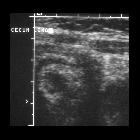
Ultrasound
Findings on ultrasound include increased symmetrical wall thickening and submucosal echogenicity. On color Doppler, there may be increased mural flow.
CT
If imaging is required, CT is usually the examination of choice.
Patients with infectious colitis from any cause typically have wall thickening (this usually demonstrates homogeneous enhancement). Low attenuation regions representing edema may be detected within the wall. Other ancillary findings include:
- ascites
- inflammation of the pericolonic fat
- multiple gas-fluid levels due to increased fluid and fluid feces
While there can be considerable overlap, the affecting segment of the portion of the colon, however, may be useful in suggesting a specific organism:
- preferentially affects the right colon (including appendix) and ileum: Yersinia, Salmonella
- diffuse involvement also occurs: cytomegalovirus, E. coli
- involvement is usually most severe in the left colon: Shigella, schistosomiasis - thought to be due to adult worms having a tendency to enter the inferior mesenteric vein
- rectosigmoid: gonorrhea, herpesvirus, and C. trachomatis (lymphogranuloma venereum)
Differential diagnosis
On imaging consider other forms of colitis dependent on the clinical situation, which includes colitis from other causes:
- inflammatory bowel disease
- ischemic colitis: usually involves watershed areas, and rarely affects the rectum
- radiation colitis
Siehe auch:


 Assoziationen und Differentialdiagnosen zu infektöse Kolitis:


