Infektionen der Milz

Computed
tomography of the spleen: how to interpret the hypodense lesion. Transverse contrast-enhanced CT images acquired during the portal-venous phase. a A 19-year-old woman with multiple pyogenic splenic abscesses during a period of immunosuppression and haematogenous spread of Staphylococcus aureus (short arrows). b A 48-year-old man with a pyogenic splenic abscess exhibiting gas formations and subcapsular fluid accumulation due to a spontaneous rupture of the abscess. c A 27-year-old man with multiple tuberculous abscesses (long arrows)

Abszess in
der Milz bei Salmonella: Beachte auch die unscharfe Kontur der Begrenzung nach ventral.

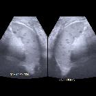
Preschooler
who is being treated for Wilms tumor and is immune suppressed and is septic. Transverse (left) and sagittal (right) US images of the spleen show multiple, round anechoic lesions within the spleen.The diagnosis was candidial microabscesses in the spleen.

Primitive
isolated hydatid cyst of the spleen: total splenectomy versus spleen saving surgical modalities. CT scan sagittal view showing a splenic hydatid cyst of 16 cm diameter

Management of
abdomen hydatidosis after rupture of a hydatid splenic cyst: a case report. CT scan demonstrating floating membrane in the splenic cyst and multiple cysts in the peritoneal cavity.

Imaging
findings of splenic emergencies: a pictorial review. Splenic infection. Axial contrast-enhanced CT of a 35-year-old man with acute myeloblastic leukemia shows low-attenuated multiple focal lesions (arrowheads) representing splenic candidiasis

Imaging
findings of splenic emergencies: a pictorial review. Splenic abscess. Axial contrast-enhanced CT of a 40-year-old woman demonstrates air (arrow) within a low attenuated splenic abscess. A culture test was positive for E.coli
Infektionen der Milz
Siehe auch:

 Assoziationen und Differentialdiagnosen zu Infektionen der Milz:

