Komplikationen bei Sinusitis

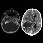
School ager
who does not take care of their teeth and had a seizure. Axial T2 MRI of the brain (above left) shows a mass in the right front lobe with surrounding vasogenic edema. Axial (above right) and coronal (below right) T1 MRI with contrast shows the mass to have thin rim enhancement and the coronal image also shows left maxillary sinusitis. The mass is shown to demonstrate diffusion restriction on diffusion-weighted imaging (below left).The diagnosis was intracranial abscess as a complication of sinusitis.

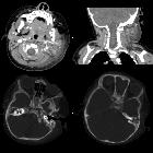
Teenager with
left orbital and forehead swelling. Sagittal CT with contrast of the brain shows frontal and left maxillary sinusitis and extensive soft tissue swelling anterior to the forehead (upper left) and destruction of the anterior left frontal bone in the frontal sinus and extensive soft tissue swelling anterior to the left orbit (upper right). Coronal (lower left) and sagittal (lower right) T1 MRI with contrast of the brain shows diffuse meningeal enhancement, subdural empyemas along the falx and both cerebral convexities, and multiple large non-enhancing subgaleal fluid collections in the left scalp.The diagnosis was meningitis, subdural empyema, and Pott puffy tumor as complications of sinusitis.
Komplikationen bei Sinusitis
Siehe auch:


 Assoziationen und Differentialdiagnosen zu Komplikationen bei Sinusitis: