Komplikationen bei Mastoiditis
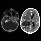
School ager
with pus draining from the right ear. Axial CT with contrast of the brain with bone windows (left) shows opacification and destruction of the right mastoid air cells while axial CT with contrast of the brain with soft tissue windows (right) shows a large low density ring enhancing lesion in the right cerebral hemisphere that is causing midline shift to the left.The diagnosis was right coalescent mastoiditis with an intracranial abscess.
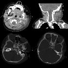
Infant with
left otitis media and left neck swelling. Axial and coronal CT with contrast of the neck with soft tissue windows (above) show extensive left cervical adenopathy and inflammation. Axial CT with contrast of the neck with bone windows (below) show bilateral complete opacification of the mastoid air cells and subtle erosive changes in the anterior aspect of the left temporal bone. There were no intracranial findings.The diagnosis was left coalescent mastoiditis.

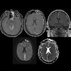
Cerebral
venous thrombosis, subdural empyema and cerebral abscess as complications of coalescent otomastoiditis. 3D MRI vascular reconstruction showing areas of hypointensity, extending from the right tranverse sinus into the sigmoid vein.

Cerebral
venous thrombosis, subdural empyema and cerebral abscess as complications of coalescent otomastoiditis. 3D T1 contrast-enhanced MRI reconstruction showing the thrombus in the right sigmoid sinus.


 Assoziationen und Differentialdiagnosen zu Komplikationen bei Mastoiditis: