Leiomyom der Harnblase


Leiomyoma of the urinary bladder is a rare benign mesenchymal tumor of the bladder. The most common presenting complaints are urinary voiding symptoms such as obstruction and irritation.
These leiomyomas exhibit imaging characteristics on ultrasound, CT and MRI similar to those of uterine leiomyomas: a well-marginated homogeneous solid mass.
Epidemiology
Leiomyomas are the most common benign bladder neoplasm but account for only 0.4% of all bladder tumors. They occur equally in men and women with a broad age range of 22-78 years . Approximately 75% of the patients are young and middle-aged .
Clinical presentation
Most are small and asymptomatic and are discovered incidentally. However, large tumors manifest with symptoms such as :
- hesitancy, frequency, dribbling
- hematuria
- pressure from mass effect
- urinary obstruction
Pathology
It is a non-infiltrative smooth muscle tumor lacking mitotic activity, cellular atypia, and necrosis. Leiomyomas arise in the submucosa, but growth may be submucosal (7%), intravesical (63%), or extravesical (30%). At cystoscopy, normal bladder mucosa covers the leiomyoma.
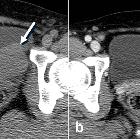
Radiographic features
Imaging features include either a smooth indentation of the bladder wall or an intraluminal mass. The lesions are smooth, solid, homogeneous masses. Cystic components indicate degeneration. The tumor exhibits characteristics similar to those of their uterine counterpart at US, CT, and MR imaging, with MR imaging being most accurate for tissue characterization.
Ultrasound
- US examination typically shows a smooth-walled homogeneous hypoechoic solid mass in the bladder with thin echogenic surface
- with US it is possible to determine the endovesical, intramural or extravesical nature of the lesion
CT
- CT is accurate in detection and localization of these lesions, and it presents as a hypoattenuating mass
- with contrast administration the leiomyoma shows moderate enhancement
MRI
MRI is superior in demonstrating the submucosal origin of the tumor and the preservation of the muscle layer. The imaging characteristics are similar to uterine leiomyomas:
- T1:
- intermediate signal intensity
- T2:
- low signal intensity
- degenerated leiomyomas have more heterogeneous signal characteristics; cystic areas have high signal intensity
- T1 C+ (Gd):
- contrast enhancement is variable; degenerated areas lack enhancement
Treatment and prognosis
Focal excision of the mass is the treatment of choice. A preoperative suspicion of a leiomyoma is invaluable in alerting the surgeon to the benign nature of the mass and preventing unnecessary radical surgery .
Differential Diagnosis
A pedunculated intraluminal leiomyoma may be confused with a urothelial lesion or urothelial cell carcinoma of the bladder but a leiomyoma should be of lower signal intensity on T2-weighted images .
Siehe auch:
- Leiomyofibrom Uterus
- Urothelkarzinom
- benigne Tumoren der Harnblase
- Neoplasien der Blase
- Leiomyosarkom der Harnblase
und weiter:


 Assoziationen und Differentialdiagnosen zu Leiomyom der Harnblase:

