Neoplasien der Blase

Multidetector
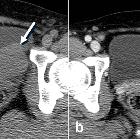
computed tomography evaluation of bladder lesions. A 57-year-old female patient with bladder leiomyoma. Axial non-contrast a and contrast-enhanced b CT images showing a smooth homogenous enhancing solid mural mass of the bladder (arrow), similar in appearance to uterine leiomyoma

Multidetector
computed tomography evaluation of bladder lesions. Urothelial (portal) and excretory (delayed) phase scans of CT urography in two patients of urothelial carcinoma. a Axial urothelial phase CT image of a 63-year-old man obtained at 70 s after bolus intravenous contrast material injection shows a small hyper-enhancing polypoid mass (arrow) that enhances more than normal bladder mucosa. b Axial excretory phase CT image shows the enhancing bladder lesion (arrow) but is less pronounced compared to the urothelial phase. c The nodule was also identified using conventional cystoscopy. d Axial urothelial phase CT image of a 74-year-old man shows an intraluminal hyper-enhancing mass (arrow) on the left wall of the bladder. e Axial excretory phase CT reveals the mass as a filling defect (arrow) within the bladder lumen. f The nodule was also detected using conventional cystoscopy

Multidetector
computed tomography evaluation of bladder lesions. Urothelial and excretory phase scans of CT urography in initial and recurred urothelial carcinomas. a Axial urothelial phase CT image of a 70-year-old man shows a small hyper-enhancing polypoid mass (arrow) that enhances more than normal bladder mucosa. b Axial excretory phase CT image shows the enhancing bladder lesion (arrow) but is less pronounced compared to the urothelial phase. c The nodule was also identified using conventional cystoscopy. Taken at the 6-month follow-up after transurethral resection of the bladder tumor, this axial urothelial phase CT image d shows an intraluminal hyper-enhancing mass (arrow) on the posterior wall of the bladder, the same as the initial tumor site. e Axial excretory phase CT of the patient shows that the mass is a filling defect (arrow) within the bladder lumen. f The nodule was detected using conventional cystoscopy

Multidetector
computed tomography evaluation of bladder lesions. CT detection of small and flat bladder lesions multiplanar reformation (MPR) in three patients of urothelial carcinoma. Axial (a) and sagittal (b) urothelial phase CT images of a 73-year-old woman showing an enhanced area of focal wall bladder thickening (arrows). Axial (c) and sagittal (d) urothelial phase CT images of a 58-year-old woman showing two tiny enhancing lesions (arrows) on the anterior wall of the bladder. Axial urothelial (e) and excretory (f) phase CT images of a 71-year-old woman showing subtle diffuse or segmental mucosal enhancement of the bladder without filling defects. The patient was diagnosed with urothelial carcinoma in situ on histopathological examination. Small or flat lesions are more difficult to detect with CT

Multidetector
computed tomography evaluation of bladder lesions. Two cases of bladder cancer. a Axial urothelial phase CT image of a 67-year-old female patient with bladder squamous cell carcinoma shows an enhancing sessile or nodular mass (arrow) in the anterior wall of the bladder. b, c Axial contrast-enhanced CT images of a 68-year-old male patient with bladder adenocarcinoma show diffuse bladder wall thickening (arrow) along the anterior wall of the bladder and a large irregular enhancing intramural mass within the bladder. There is irregular soft tissue stranding (small arrow) from tumor invasion into the perivesical fat

Multidetector
computed tomography evaluation of bladder lesions. A 67-year-old male patient with bladder leiomyosarcoma. Axial (a), coronal (b), and sagittal (c) contrast-enhanced CT images showing a large, poorly circumscribed, heterogeneously enhancing mass (arrows) on the dome of the bladder wall

Multidetector
computed tomography evaluation of bladder lesions. A 60-year-old male patient with bladder lymphoma. Axial contrast-enhanced CT image showing marked circumferential wall thickening of the bladder (arrow)

Multidetector
computed tomography evaluation of bladder lesions. Two cases of bladder papilloma and papillary urothelial neoplasm of low malignant potential (PUNLMP). Axial urothelial phase a CT image of a 60-year-old male patient with bladder papilloma showing a tiny enhancing lesion (arrow) on the trigone of the bladder and also demonstrating a filling defect (arrow) on the excretory phase (b) CT image. Axial urothelial phase c of a 19-year-old male patient with PUNLMP and excretory phase (d) CT images showing an irregular polypoid enhancing lesion and filling defect (arrows) in the bladder mimicking bladder cancer
Neoplasien der Blase
Siehe auch:
- Harnblasenkarzinom
- Urachuskarzinom
- benigne Tumoren der Harnblase
- Harnblasenpolyp
- Plattenepithelkarzinom der Harnblase
- Lymphom Harnblase
- verkalkter Tumor der Blase
- Blasentumoren bei Kindern
- Osteosarkom der Blase
- tumor-like lesions of the urinary bladder
- Rhabdomyosarkom der Blase
- angiosarcoma of the urinary bladder
und weiter:

 Assoziationen und Differentialdiagnosen zu Neoplasien der Blase:


