Harnblasenkarzinom

Advanced
squamocellular HIV-related urinary bladder carcinoma. Portal venous phase post-contrast images (b,c) confirmed urinary bladder almost entirely replaced by large (over 10 cm) mass with markedly heterogeneous, necrotic-like enhancement.

Advanced
squamocellular HIV-related urinary bladder carcinoma. Portal venous phase post-contrast images (b,c) confirmed urinary bladder almost entirely replaced by large (over 10 cm) mass with markedly heterogeneous, necrotic-like enhancement.

Advanced
squamocellular HIV-related urinary bladder carcinoma. Excretory phase post-contrast images (d...f) confirmed urinary bladder almost entirely replaced by large (over 10 cm) mass with markedly heterogeneous, necrotic-like enhancement. Note patent, non-dilated distal ureters (arrows).

Advanced
squamocellular HIV-related urinary bladder carcinoma. Excretory phase post-contrast images (d...f) confirmed urinary bladder almost entirely replaced by large (over 10 cm) mass with markedly heterogeneous, necrotic-like enhancement. Note patent, non-dilated distal ureters (arrows).

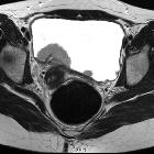
Advanced
squamocellular HIV-related urinary bladder carcinoma. Multiplanar T2-weighted images (a..c) confirmed severe (over 3 cm) diffuse mural thickening (*) of the urinary bladder with solid intermediate signal intensity, consistent with full-thickness tumour.

Advanced
squamocellular HIV-related urinary bladder carcinoma. Multiplanar T2-weighted images (a..c) confirmed severe (over 3 cm) diffuse mural thickening (*) of the urinary bladder with solid intermediate signal intensity, consistent with full-thickness tumour.

Advanced
squamocellular HIV-related urinary bladder carcinoma. Multiplanar T2-weighted images (a..c) confirmed severe (over 3 cm) diffuse mural thickening (*) of the urinary bladder with solid intermediate signal intensity, consistent with full-thickness tumour. Note infiltration of perivesical fat (arrowheads).

Advanced
squamocellular HIV-related urinary bladder carcinoma. The severe (over 3 cm) diffuse mural thickening (*) of the urinary bladder showed solid T1 signal intensity. Note infiltration of perivesical fat (arrowheads), minimal residual lumen (+).

Advanced
squamocellular HIV-related urinary bladder carcinoma. After intravenous gadolinium, the neoplastic mural thickening of the urinary bladder showed markedly heterogeneous contrast enhancement. Note infiltration of perivesical fat (arrowheads), opacified residual lumen (+).

Advanced
squamocellular HIV-related urinary bladder carcinoma. After intravenous gadolinium, the neoplastic mural thickening of the urinary bladder showed markedly heterogeneous contrast enhancement. Note infiltration of perivesical fat (arrowheads), opacified residual lumen (+).

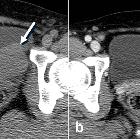
Multidetector
computed tomography evaluation of bladder lesions. Two cases of bladder cancer. a Axial urothelial phase CT image of a 67-year-old female patient with bladder squamous cell carcinoma shows an enhancing sessile or nodular mass (arrow) in the anterior wall of the bladder. b, c Axial contrast-enhanced CT images of a 68-year-old male patient with bladder adenocarcinoma show diffuse bladder wall thickening (arrow) along the anterior wall of the bladder and a large irregular enhancing intramural mass within the bladder. There is irregular soft tissue stranding (small arrow) from tumor invasion into the perivesical fat
Transitional
cell carcinoma (urinary tract) • Urothelial cell carcinoma - stage T3b - Ganzer Fall bei Radiopaedia

Small cell
carcinoma (urinary bladder) • Small cell carcinoma of the bladder - Ganzer Fall bei Radiopaedia

Bladder
cancer • Urachal carcinoma - Ganzer Fall bei Radiopaedia

Bladder
cancer • Locally invasive urinary bladder cancer - Ganzer Fall bei Radiopaedia
Bladder cancer is a broad term used to describe all types of cancers affecting the urinary bladder:
- transitional cell carcinoma (urinary bladder): most common primary neoplasm of the bladder
- squamous cell carcinoma (urinary bladder): accounts for around 3-8% of all bladder cancers
- adenocarcinoma (urinary bladder): accounts for around 1% of all bladder cancers
- small cell carcinoma (urinary bladder): extremely rare
Staging
The Vesical Imaging-Reporting and Data System (VI-RADS) scoring system was created in 2018 to standardize imaging and reporting of bladder cancer staging with multiparametric MRI .
Siehe auch:
- Urothelkarzinom
- Urachuskarzinom
- Neoplasien der Blase
- Plattenepithelkarzinom der Harnblase
- Lymphom Harnblase
- Blasentumoren bei Kindern
- Transitionalzellkarzinom der Blase
- Rhabdomyosarkom der Blase
und weiter:


 Assoziationen und Differentialdiagnosen zu Harnblasenkarzinom: