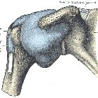
Ligamentum coracohumerale

The left
shoulder and acromioclavicular joints, and the proper ligaments of the scapula.

MR
arthrography of SLAP X tear with rotator interval tear and biceps tendon rupture. Coronal images comparing a torn coracohumeral ligament in this patient with a normal study.

Alteration in
coracohumeral ligament and distance in people with symptoms of subcoracoid impingement. The illustration of USG measurement. A. coracohumeral ligament (CHL) B. a. coracohumeral distance (CHD) in external rotation (ER), b. neutral rotation (NR), c. internal rotation (IR) and d. internal rotation with maximal flexion and adduction (IRFA). CP: coracoid process HH: humerus head, C: coracoid process HH: humerus head

Accuracy of
unguided and ultrasound guided Coracohumeral ligament infiltrations – a feasibility cadaveric case series. Ultrasound scan guided Coracohumeral ligament periligamentous injection of a right shoulder showing coracohumeral ligament: white arrow heads; HH: Humeral head; CP: Coracoid process; injecting needle: blue arrow heads. Transducer placement over the anterior superior aspect of the shoulder, with the coracohumeral ligament in the long axis
The coracohumeral ligament (CHL) is a strong supportive ligament of the shoulder joint and is a part of the rotator cuff interval.
Gross anatomy
- originates from the lateral surface of the base of the coracoid process
- runs laterally across the glenohumeral capsule and covers the long head of the biceps tendon superiorly
- attaches to the margin of the greater and lesser tubercles of the humerus, and along the transverse ligament bridging the bicipital groove
Variant anatomy
- rarely hypoplastic/aplastic (~5%)
Siehe auch:
- Ligamentum coracoclaviculare
- Ligamentum transversum humeri
- Ligamentum coracoacromiale
- Pulleyläsion
- subcoracoidales Impingement
und weiter:

 Assoziationen und Differentialdiagnosen zu Ligamentum coracohumerale:


