Lobus venae azygos


An azygos lobe is created when a laterally displaced azygos vein creates a deep pleural fissure into the apical segment of the right upper lobe during embryological development. It is a normal anatomic variant of the right upper lobe due to the invagination of the azygos vein and pleura during development in the fetus. It is not a true accessory lobe as it does not have its own bronchus.
Epidemiology
An azygos lobe is found in 1% of anatomical specimens and is twice as common in males as females .
Embryology
An azygos lobe forms when the right posterior cardinal vein, one of the precursors of the azygos vein, fails to migrate over the apex of the lung and penetrates it instead, carrying along with two pleural layers that invaginate into the upper portion of the right upper lobe .
Radiographic features
Plain radiograph
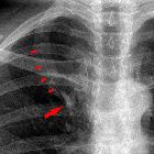
The azygos lobe is usually well seen on the chest radiograph, where it is limited by the azygos fissure, a fine, convex (relative to the mediastinum) line that crosses the apex of the right lung.
CT
CT shows the deep penetration of the lung behind the SVC and the trachea.
The azygos fissure extends from the lateral aspect of the vertebral body posteriorly, to the right brachiocephalic vein and SVC anteriorly. This consists of two layers of parietal pleura and two layers of visceral pleura .
The azygos vein is seen as a thicker structure following the same path as the fissure. The position of the arch is higher than when it follows an intramediastinal course
The azygos vein usually ends in the SVC, but occasionally in the right brachiocephalic vein.
Siehe auch:
- akzessorische Lungenlappen
- Vena azygos
- enlarged azygos vein in an azygos fissure from azygos continuation of IVC
und weiter:


 Assoziationen und Differentialdiagnosen zu Lobus venae azygos: