Lungenmetastasen Wilmstumor

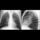
Preschooler
with Wilms tumor. CXR PA shows a large round opacity just lateral to the left pulmonary artery which is located anteriorly on the lateral view.The diagnosis was a lung metastasis in a patient with Wilms tumor.

Wilms tumoru
with bony-mandibular, rib and calvarial metastases. Another lung parenchymal lesion seen adjacent to the pleural-based soft tissue.

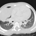
Teenager with
past history of Wilms tumor. CXR AP (above) shows a soft tissue density projecting in the right cardiophrenic angle. Axial CT with contrast of the chest (below) shows a soft tissue mass in the right posterior costophrenic sulcus.The diagnosis was lung metastasis in a patient with Wilms tumor.

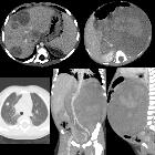
Toddler with
abdominal pain and distension, hematuria and lethargy for 2 months. Axial (above right), coronal (below middle) and sagittal (below right) CT with contrast of the abdomen shows a large heterogenous non-calcified mass that fills the entire left side of the abdomen. The inferior vena cava (to the right of the aorta) was distended with tumor thrombus. Multiple liver (above left) and lung (below left) lesions are also seen.The diagnosis was Wilms tumor with liver metastases and lung metastases and invasion of the inferior vena cava.
Lungenmetastasen Wilmstumor
Siehe auch:

 Assoziationen und Differentialdiagnosen zu Lungenmetastasen Wilmstumor:

