lupus mastitis

Recurrent
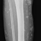
breast lesion in a patient with systemic lupus erythematosus – A radiological review and case report. 1.5 Tesla MR, T2 image showing a well-defined, inhomogeneous mass on the right upper quadrant.

Recurrent
breast lesion in a patient with systemic lupus erythematosus – A radiological review and case report. Mammography showing a focal inhomogeneous hyperdensity with calcification clusters.

Recurrent
breast lesion in a patient with systemic lupus erythematosus – A radiological review and case report. Sonography showing an ill-defined hypoechogenicity with associated calcifications on the right upper quadrant.

Recurrent
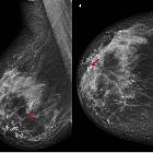
breast lesion in a patient with systemic lupus erythematosus – A radiological review and case report. Recurrent spiculated inhomogeneous breast mass with microcalcifications on the 18-month follow-up after initial surgical resection.
Lupus
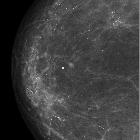
Mastitis - rare but typical imaging findings. Oblique lateral (A) and craniocaudal mamography incidences (B) show coarse and diffuse calcifications (red arrows).

Lupus
Mastitis - rare but typical imaging findings. Oblique lateral (A) and craniocaudal mamography incidences (B) represent the previous exam, 2 years before the mamography shown in figure 1. Note that in this exam there are much less calcifications.

Lupus
Mastitis - rare but typical imaging findings. Breast ultrasound shows fibrotic changes in the glandular tissue and a coarse calcification (red arrow).


 Assoziationen und Differentialdiagnosen zu Systemischer Lupus Erythematodes (Manifestationen in der Mamma):