malignes Mesotheliom

Mesotheliom
der Pleura links in der Computertomographie: Mantelförmige Verdickung der Pleura mit Pleuraerguss. Beachte vor allem auch die Ausbreitung an der mediastinalen Seite der Pleura.

Mesotheliom
der Pleura links in der Computertomographie: Mantelförmige Verdickung der Pleura mit Pleuraerguss. Beachte vor allem auch die Ausbreitung an der mediastinalen Seite der Pleura.

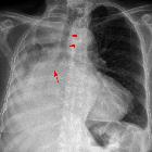
Pleuramesotheliom
im Röntgenbild des Thorax: Pleuraverdickung und -erguß links (rechts im Bild).

This image
is part of a series which can be scrolled interactively with the mousewheel or mouse dragging. This is done by using Template:Imagestack. The series is found in the category Peritoneal mesothelioma - CT - case 001. Peritoneales desmoplastisches Mesotheliom in der Computertomographie. Dieses Bild ist Teil einer Serie zum Durchblättern (siehe Kategorie).

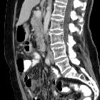
CT -
gesteuerte Biopsie einer zu diesem Zeitpunkt unklaren peritonealen Tumorausbreitung mit Verdacht auf Peritonealkarzinose ohne Nachweis eines Primarius. Histologie: Peritoneales Mesotheliom.

Mesotheliom
der Pleura links in der Computertomographie: Mantelförmige Verdickung der Pleura mit Pleuraerguss. Beachte vor allem auch die Ausbreitung an der mediastinalen Seite der Pleura.

Mesotheliom
der Pleura rechts in der Computertomographie: Mantelförmige Verdickung der Pleura. Ein Pleuraerguss war basal auch vorhanden. Beachte auch die deutliche Volumenminderung der betroffenen Seite.

Pleuramesotheliom
rechts: Zirkuläre Verdickung der Pleura. Volumenminderung der rechten Lunge.

Pleuramesotheliom
im Röntgenbild: Zirkuläre Verbreiterung der Pleura links mit Pleuraerguss.


 Assoziationen und Differentialdiagnosen zu malignes Mesotheliom: