solitärer fibröser Tumor der Pleura


Solitary fibrous tumor of the pleura, also known as pleural fibroma, is a rare benign pleural-based tumor that accounts for <5% of all tumors involving the pleura.
Epidemiology
Usually presents in the 6 to 7 decades. There is no recognized gender predilection. It has an incidence of 2.8 per 100,000 persons .
Clinical presentation
Usually asymptomatic and discovered as an incidental finding on a routine chest radiograph . Of those who are symptomatic, the clinical presentation can be with either a cough, chest pain, or shortness of breath.
Associations
- hypoglycemia (2-4%)
- thought to be due to the production of insulin-like growth factor 2
- hypertrophic pulmonary osteoarthropathy (~20%)
- thought to be due to abnormal production of hyaluronic acid
Asbestos exposure is not an association.
Pathology
They are composed of irregularly arranged fascicles consisting of spindle cells separated by collagen. Thought to originate from submesothelial mesenchymal cells. Approximately 80% of pleural fibromas arise from the visceral pleura. Myxoid or cystic degeneration can occur.
Location
There may be a predilection towards the mid to lower zones of the chest. In ~80% of cases, they arise from visceral pleura, with the remainder arising from the parietal pleura.
Variants
- lipomatous hemangiopericytoma: a fat-forming variant of pleural fibroma
Radiographic features
Plain radiograph
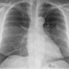
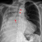
Presents as a pleural-based mass, which tends to be relatively circumscribed, but can also be lobulated. It often forms an obtuse angle with the chest wall and may grow to a large size. Pedunculated lesions can change position and appearance with respiration or with a change in position on serial radiographs.
Calcification, rib destruction, and pleural effusions are typically not associated features.
CT
Tends to have soft tissue attenuation on unenhanced scans and shows relatively homogenous intense background enhancement on contrast-enhanced scans (from rich vascularization). Non-enhancing areas may be present, corresponding to necrosis, myxoid degeneration, or hemorrhage within the tumor. A pedicular attachment may also be seen.
MRI
Due to the fibrous component, signal characteristics tend to be:
- T1: typically low to intermediate signal
- T2: typically low signal overall (thought to be due to high cellularity and abundant collagen); areas of necrosis and myxoid degeneration can have high signal
MR may also show necrotic, hemorrhagic, and cystic components in greater detail if these are present.
Treatment and prognosis
The majority of tumors tend to be benign and slow-growing. Malignant tumors are uncommon but have been reported. Surgical resection is the treatment of choice . There can be a recurrence in a small proportion (8%) of cases, most of which do not recur again following re-excision.
History and etymology
Understanding of primary pleural neoplasia has changed drastically since the late 1800s, and so the associated terminology has also changed. German pathologist Ernst Leberecht Wagner (1829-1888)is generally credited with the first description of pleural mesothelioma / pleural fibroma in 1870, although he described it "tuberkelähnliche" lymphadenome ("tubercle-like" lymphadenoma) . Primary pleural malignancies were rare until the twentieth century when incidence increased considerably due to environmental exposures.
Throughout much of the twentieth century, there was a continual debate about the precise histology of both normal pleura and pleural tumors. The properties of the mesothelial cell itself were controversial, mostly due to its unique expression of both mesenchymal and epithelial attributes.
Gradually, a distinction was recognized between "localized" versus aggressive mesothelial tumors (i.e. "malignant" mesothelioma). The former entity has been variably termed benign mesothelioma, localized fibrous tumor of the pleura, localized mesothelioma, localized fibrous mesothelioma, localized benign fibroma, or submesothelial fibroma .
Differential diagnosis
Considerations for extremely well-defined lesions include:
- pleural lipoma
- intercostal nerve neurilemoma (schwannoma)
If not extremely well defined, consider:
- organized inflammation
- peripheral bronchogenic carcinoma
- solitary pleural metastasis
Also, consider the differential for a single pleural mass.
Siehe auch:
- pleurale Lipome
- Lungenkarzinom
- hypertrophe Osteoarthropathie
- benigne pleurale Tumoren
- solitärer fibröser Tumor
- einzelne Pleuraraumforderung
- Tumoren der Pleura
- Doege-Potter-Syndrom
- maligner solitärer fibröser Tumor der Pleura
- Hypoglykämie
und weiter:


 Assoziationen und Differentialdiagnosen zu solitärer fibröser Tumor der Pleura: