Single pleural based mass (differential)

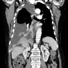
This axial CT
image with intravenous contrast reveals what appears to be a posterior mediastinal mass, which was surgically removed and found to be a solitary fibrous tumor of the pleura.

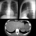
Lipom der
Pleura links dorsal in der Computertomographie. Oben im Lungenfenster, unten im Weichteilfenster, dort aufgrund der geringen Dichte fast zu übersehen. Zufallsbefund im Röntgen-Thorax.

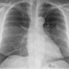
This PA chest
radiograph demonstrates an abnormal contour in the right hilar region, with visualization of the pulmonary vessels through the mass (the hilar overlay sign) indicating its posterior mediastinal location. On resection this was found to be a benign solitary fibrous tumor of the pleura.


The differential for a single pleural mass is essentially the same as that for multiple pleural masses with the addition of a few entities.
- tumors
- pleural tumors
- metastatic pleural disease, particularly from adenocarcinomas, e.g.
- bronchogenic adenocarcinoma
- breast cancer
- ovarian cancer
- prostate cancer
- gastrointestinal adenocarcinoma
- renal cell carcinoma
- lymphoma: pleural lymphoma
- invasive thymoma
- lipoma
- loculated fluid (on plain film)
- mass related to ribs or chest wall, e.g. Ewing sarcoma of chest wall, Askin tumor
- mass related to the intercostal nerve
- splenosis - thoracic splenosis
- infection including tuberculosis
Siehe auch:
- Tuberkulose
- pleurale Lipome
- Pleuraerguss
- Pleuraempyem
- Pleurakarzinose
- Empyem
- Splenose
- solitärer fibröser Tumor der Pleura
- benigne pleurale Tumoren
- Pleuramesotheliom
- Adenokarzinom der Prostata
- maligne pleurale Tumoren
- invasives Thymom
- pleurale Metastasen
- pleurale Lipomatose
- localised mediastinal malignant mesothelioma
- extrapleuraler solitärer fibröser Tumor
- Askintumor
- multiple pleural masses
und weiter:

 Assoziationen und Differentialdiagnosen zu einzelne Pleuraraumforderung:


