maligne pleurale Tumoren

Malignant
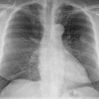
solitary fibrous tumor of the pleura slowly growing over 17 years: case report. Serial chest radiographs from 1995 through 2012. (A) The triangular-shaped mass in the right pleural cavity was identified. (B) The mass was stable. (C) The mass exhibited progression in 2006. (D) The mass had grown significantly.

Primary
neoplasms of the thymus • Thymoma - invasive - Ganzer Fall bei Radiopaedia

Pleural
synovial sarcoma. Expiratory chest radiograph demonstrates a moderate to large left pleural effusion with superimposed consolidation, initially interpreted as pneumonia with parapneumonic effusion. There is also a rounded opacity in the left suprahilar region, concerning for mass.

Pleural
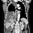
synovial sarcoma. Coronal (A) and axial (b) non-contrasted chest CT images demonstrate a rounded posterior mediastinal mass with associated left pleural effusion and left lower lobe atelectasis.

Malignant
pleural disease • Pleural synovial sarcoma - Ganzer Fall bei Radiopaedia

Malignant
pleural disease • Pleural malignancy (pleural epithelioid angiosarcoma) - Ganzer Fall bei Radiopaedia

Malignant
pleural disease • Pleural effusion - unilateral - malignant - Ganzer Fall bei Radiopaedia

Malignant
pleural disease • Pleural metastases - Ganzer Fall bei Radiopaedia

Malignant
pleural disease • Pleural metastases from melanoma - Ganzer Fall bei Radiopaedia

Non-Hodgkin
lymphoma • Malignant pleural thickening - non-Hodgkin lymphoma - Ganzer Fall bei Radiopaedia

Malignant
pleural disease • Pleural carcinomatosis - Ganzer Fall bei Radiopaedia

Malignant
pleural disease • Pleural metastases - thyroid cancer - Ganzer Fall bei Radiopaedia

Asbestos-related
diseases • Mesothelioma - Ganzer Fall bei Radiopaedia

Multidetector
CT findings of primary pleural angiosarcoma : a systematic review, an additional cases report. Enhanced multidetector CT (A, B) of a 67-year-old man showed right circumscribed, necrotic pleural masses. PET-CT(C) showed very intense uptake at the right pleural masses. 2D enhanced coronal reformat image (D) showed right visceral (dashed arrows) and parietal pleural masses(arrows)

Pleuramesotheliom
im Röntgenbild: Zirkuläre Verbreiterung der Pleura links mit Pleuraerguss.

Multidetector
CT findings of primary pleural angiosarcoma : a systematic review, an additional cases report. Primary pleural angiosarcoma in a 75-year-old man with a history of tuberculosis. Enhanced multidetector CT scan (A, B) showed two, sharply demarcated, biconvex, hypodense masses, arising from chronic tuberculous empyema sequelae with calcific plaques. These masses were directly invaded the extrathoracic regions, resulting in adjacent left 6-8th ribs destruction (B, C). Lung scan (D) CT showed multiple hematogenous metastases in both lung fields, and left rib posterior arc change

Multidetector
CT findings of primary pleural angiosarcoma : a systematic review, an additional cases report. A 65-year-old man diagnosed with primary pleural angiosarcoma. Enhanced, multidetector chest CT scans showed multiple, small nodules of poorly enhancing nature in the left parietal pleura(asterisk) (A). There were also right large pleural effusion and left small pleural effusion with thin, parietal pleural enhancement (B, C)

Tumor and
tumorlike conditions of the pleura and juxtapleural region: review of imaging findings. Diagnosis: angiosarcoma. Technique: contrast-enhanced chest CT. Description: A 64-year-old man with no history presented with progressive shortness of breath. The initial contrast-enhanced CT scan (a) to rule out pulmonary embolism shows a well-delineated pleural-based oval lesion (arrow) in the base of the right lung. Findings are relatively aspecific. Follow-up CT examination 4 weeks later (b) shows prominent disease progression, with a large amount of fluid. Despite the relatively short time interval, there is a marked increase in pleural lesions, with widespread focal contrast enhancing pleural lesions in the right hemithorax. The rapid and aggressive progression points to the possible diagnosis of angiosarcoma, which was histologically confirmed

Malignant
solitary fibrous tumor of the pleura slowly growing over 17 years: case report. Pre-operative imaging finding. (A) Computed tomography of the chest revealed a large mass in the right pleural cavity, which was inhomogeneous with peripheral enhancement. (B) Positron emission tomography-CT also showed a pleural mass with low metabolic activity (standardized uptake value: 3.17).

Desmoplastic
round cell tumour of pleura with liver and spine metastases: Uncommon pathology with grave prognosis. Coronal reformation demonstrates multiple hypodense lesions in the lower thoracic spine (metastasis) with heterogeneously enhancing nodular thickening on LT side causing collapse of underlying lung parenchyma.

A case of
malignant solitary fibrous tumour of the mediastinal pleura. Coronal chest CT image shows a well-circumscribed round enhancing solid mass located in the middle part of the mediastinum, with eccentric calcification (arrow).

A case of
malignant solitary fibrous tumour of the mediastinal pleura. Coronal chest CT image shows a well-circumscribed round mild heterogeneous enhancing solid mass located in the middle part of the mediastinum, with left dislocation of the oesophagus (arrows).

A case of
malignant solitary fibrous tumour of the mediastinal pleura. Axial chest CT image shows a well-circumscribed round enhancing solid mass located in the middle part of the mediastinum.

Pleuramesotheliom
rechts: Zirkuläre Verdickung der Pleura. Volumenminderung der rechten Lunge.


Mesotheliom
der Pleura rechts in der Computertomographie: Mantelförmige Verdickung der Pleura. Ein Pleuraerguss war basal auch vorhanden. Beachte auch die deutliche Volumenminderung der betroffenen Seite.

Pleural
synovial sarcoma. Coronal (A) and axial (b) non-contrasted chest CT images demonstrate a rounded posterior mediastinal mass with associated left pleural effusion and left lower lobe atelectasis.
maligne pleurale Tumoren
pleurale Tumoren Radiopaedia • CC-by-nc-sa 3.0 • de
There are several tumors that can involve the pleura which can range from being benign to malignant. The list includes:
- primary pleural tumors
- mesothelial tumors
- pleural malignant mesothelioma
- well-differentiated papillary mesothelioma
- adenomatoid tumor
- mesenchymal tumors
- solitary fibrous tumor (pleural fibroma)
- pleural angiosarcoma
- pleural synovial sarcoma
- desmoid-type fibromatosis
- calcifying fibrous tumor
- desmoplastic round cell tumor
- lymphoproliferative disorders
- mesothelial tumors
- secondary lesions that can involve the pleura
- metastases: see pleural metastases
- lung cancer
- breast carcinoma
- ovarian cancer
- lymphoma
- gastric carcinoma
- thymic epithelial tumors (thymoma)
- invasive tumors to the pleura
- thymic epithelial tumors (thymoma) with pleural invasion
- pericardial tumors with pleural invasion
- invasive chest wall tumors
- Ewing sarcoma family of tumors with pleural invasion
- metastases: see pleural metastases
See also
- single pleural mass: differential
- tumor-like conditions of the pleura
Siehe auch:
- Pleuraerguss
- Pleurakarzinose
- benigne pleurale Tumoren
- Pleuramesotheliom
- pleurale Tuberkulose
- einzelne Pleuraraumforderung
- Tumoren der Pleura
- pleurale Metastasen
- extraossäres Plasmozytom
- Chondrosarkom der Pleura
- Synovialsarkom der Pleura
- mesothelioma-like pleuropulmonary angiosarcoma
- desmoplastischer klein- und rundzelliger Tumor (DSRCT) der Pleura
- maligner solitärer fibröser Tumor der Pleura
- noduläre Pleuraverdickung
und weiter:

 Assoziationen und Differentialdiagnosen zu maligne pleurale Tumoren:


