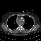
Mediastinalhämatom

A diagnostic
approach to the mediastinal masses. a Contrast-enhanced CT scan of a man who had suffered a traffic accident. An infiltrative mediastinal haematoma is identified with subtle areas of high CT-attenuation values (arrow). Bilateral pleural effusion (*) and a sternum fracture (open arrow) are also observed. b Iatrogenic mediastinal haematoma (arrow) in a 64-year-old man secondary to bronchoscopy with transtracheal biopsy. Note the high attenuation value of the lesion compared with muscular tissue

A case of
mediastinal haematoma mimicking a mediastinal mass. Arterial phase coronal reconstruction shows no increase in HU values. No cystic density, fat attenuation, calcifications, tracheal deviation, vascular compression.
Posterior
mediastinal hematoma – a rare case following a fall from standing height: a case report. Posterior Mediastinal hematoma-high mediastinum – Please note the compressed trachea.

A surgical
case of mediastinal hematoma caused by a minor traffic injury. CT findings. a A cervical hematoma in the retropharyngeal space. b Mediastinal hematoma. c A mediastinal hematoma compressing the trachea, esophagus, and superior vena cava. d Extravasation of contrast medium (arrow)


 Assoziationen und Differentialdiagnosen zu Mediastinalhämatom:


