Milzarterienaneurysma


Splenic artery aneurysms are the commonest visceral arterial aneurysm formation as well as the 3 commonest abdominal aneurysm (after the aorta and iliac vessels). Aneurysms are usually saccular in configuration and they can either be in the form of a true aneurysm (much more common) or as a pseudoaneurysm.
This article focus on the true splenic artery aneurysm, please refer on splenic artery pseudoaneurysms for a specific discussion on this entity.
Epidemiology
- the true prevalence of splenic artery aneurysm is unknown.
- estimates vary widely from 0.2% to 10.4%, but generally it is the third most common site of intra-abdominal aneurysms after abdominal aorta and iliac arteries .
- incidentally discovered splenic artery aneurysms are being diagnosed more frequent with wider use of cross-sectional imaging modalities .
- a splenic artery aneurysm is about four times more common in females, yet the risk of its rupture is about three times more common in males .
Associations
Clinical presentation
Most splenic artery aneurysms are silent and are discovered in asymptomatic patients . More than 95% of patients with non-ruptured splenic artery aneurysms were asymptomatic . Risk of rupture increases with liver transplantation, portal hypertension, and pregnancy. A ruptured splenic artery aneurysm usually presents with sudden onset left upper quadrant abdominal pain followed by hemodynamic instability, and gastrointestinal bleeding .
The so-called “double rupture” phenomenon occurs when initial spontaneous stabilization followed by subsequent sudden circulatory collapse is experienced. This is caused by initial bleeding collecting into the lesser sac then followed by flooding into the peritoneal cavity .
Radiographic features
General
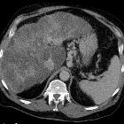
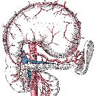
Size of splenic artery aneurysms can range from 2 to 9 cm, but usually it is smaller than 3 cm. Those may be single or multiple and are most commonly involving the distal portion of the artery. Peripheral calcification is common, and mural thrombus may be present .
Several imaging modalities can be used to diagnose splenic artery aneurysms.
Ultrasound
Ultrasound with Doppler is non ionizing modality and relatively cheap and available yet it is operator dependent, has limited spatial resolution and may be difficult in cases of obesity, bowel shadowing and atherosclerosis.
MR angiography
MR angiography is of higher spatial resolution than US but it is relatively expensive and contraindicated in some patients (claustrophobia, aneurysm clips, cardiac pacemakers).
CT angiography
CT angiography is a quick and efficient examination, has higher resolution than MR and its MPR capabilities greatly help in diagnosis, yet it has its contraindications (renal failure, allergy to contrast).
Direct catheter angiography
Direct catheter angiography is the gold standard imaging test, has the highest resolution and concomitant management can be applied, yet it still carries the complications of arterial puncture .
Treatment and prognosis
The overall risk of rupture is thought to be ~6%. However in an event of rupture there is a relatively high mortality rate of ~36% .
Follow-up of incidentally-detected splenic artery aneurysms :
- <2 cm
- spontaneous rupture is rare
- 1 year follow up, if no risk factors
- follow up interval may be extended if other comorbidities are present, or if there is a decreased life expectancy
- spontaneous rupture is rare
- ≥2 cm
- endovascular therapy should be considered
- coil embolization is increasingly used to treat larger aneurysms
- endovascular therapy should be considered
Rapidly increasing size, presence in a premenopausal woman, cirrhosis, and symptomatic aneurysm may warrant intervention, regardless of size.
Differential diagnosis
- if only limited views of the upper abdomen are available, it may be difficult to differentiate from a (calcified) left adrenal hematoma
See also
Siehe auch:
- Leberzirrhose
- Vaskulitis
- Fibromuskuläre Dysplasie
- portale Hypertension
- Arteria lienalis
- verkalktes Milzarterienaneurysma
- Atherosklerose
- Pseudoaneurysma der Milzarterie
- multiple Aneurysmen der Milzarterie
- rupturiertes Milzarterienaneurysma
und weiter:


 Assoziationen und Differentialdiagnosen zu Milzarterienaneurysma: