mucoepidermoid carcinoma (MEC) of lung


Mucoepidermoid carcinoma (MEC) of the lung is a type of non small cell lung cancer (NSCLC). It is classified under the group of lung carcinomas of the salivary gland type.
Epidemiology
Mucoepidermoid carcinoma (MEC) is the most common of the SGTTLs . The tumor is thought to account for ~ 0.1-0.2% of primary lung cancers . MEC may be encountered in any age group, however, most cases have been seen in adults . There appears to be slight male preponderance with a male-to-female distribution of almost 1.5:1 .
Pathology
Lesions typically occur in relation to the tracheobronchial tree (hence they are also termed muco-epidermoid carcinomas of the tracheobronchial tree). In larger series, no topographic predilection for any particular pulmonary lobe or segment has been discovered. Central location often leads to postobstructive mucoid and lipoid pneumonia .
Gross pathology
Common presentation of this uncommon tumors may be as exophytic endobronchial tumor, potentially greater than 5 cm in greatest diameter. Usually well circumscribed and with smooth overlying mucosal surfaces, their cut sections are tan-gray or yellow.
Histopathology
May be solid, cystic or both, may show overtly mucoid features. Components comprise clear cells, squamoid cells or transitional polygonal cells, interspersed with areas containing mucus-secreting glandular cells.
They are divided into low- and high-grade lesions:
- low-grade tumors characteristically demonstrate bland cytologic features; mitotic activity is minimal or absent
- high-grade mucoepidermoid carcinoma demonstrates a greater degree of cytologic anaplasia in both its squamoid and glandular elements; areas of necrosis and hemorrhage may also be present
Differentials on biopsy
- low-grade MEC
- may resemble mucous gland adenoma (MGA), especially in small biopsies
- distinction between these two entities may be impossible without complete resection of the tumor
- high-grade MEC: distinction from NSCLC may be largely academic
- usually based on the absence of foci of conventional adenocarcinoma
- other usable features include absence of an in situ carcinomatous component and the presence of low-grade mucoepidermoid areas in some of these high-grade lesions
Clinical presentation
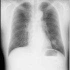
Symptoms are dependent on the size and location of the neoplasms. Large central tumors can cause symptoms of obstruction, with pneumonia, dyspnea or chest pain, whilst more peripheral lesions may be asymptomatic. In these cases, they are frequently encountered on routine chest X-ray.
Radiographic features
CT
CT features are can be variable and nonspecific, although a well-defined ovoid or lobulated intraluminal or lung peripheral mass with moderate to marked heterogeneous contrast enhancement may suggest towards the diagnosis .
Lesions may sometimes show punctate calcification and may adapt to branching feature of the airways .
FDG PET/CT
May aid in differentiation from other lung tumors , but first of all, it has been shown to have high accuracy in detection of histopathological tumor differentiation :
- high-grade - high FDG-hypermetabolism (avid)
- low-grade - slight FDG-hypermetabolism (less or even non avid)
Hence prediction of prognosis (and need for more aggressive treatment) may be possible .
Treatment and prognosis
Most lesions are generally regarded as low grade and overall prognosis may be more favorable than other forms of lung cancer.
For high-grade tumors prognosis may equate that of other forms of NSCLC s , hence adjuvant radiation and chemotherapy is often performed .
Siehe auch:
- Tumoren der Trachea
- Tumoren des Tracheobronchialsystems
- mukoepidermoides Karzinom
- Lungenkarzinome vom Speicheldrüsentyp
- mukoepidermoides Karzinom des Tracheobronchialsystems
und weiter:


 Assoziationen und Differentialdiagnosen zu mukoepidermoides Karzinom der Lunge:


