Lungenkarzinome vom Speicheldrüsentyp

Mucoepidermoid
carcinoma of lung • Bronchial mucoepidermoid carcinoma - Ganzer Fall bei Radiopaedia

Mucoepidermoid
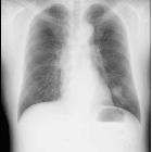
carcinoma of the lung: a case report. Chest radiography revealed a mass shadow in the left lower lung field.

Mucoepidermoid
carcinoma of the lung: a case report. CT showed a mass shadow measuring 30 mm in diameter in S9.

Bone marrow
metastasis in primary bronchial mucoepidermoid carcinoma: a case report. Enhanced CT scan revealing two lobulated masses measuring 30 to 40 mm in diameter in the left inferior lobar bronchus (A) and peripheral tissue of the lower lobe of the left lung (B), respectively.

Right
pneumonectomy for primary large acinic cell carcinoma (AciCC) with severe mediastinal deviation: a case report and literature review. Preoperative imaging of the large acinic cell carcinoma of right lung. A Preoperative chest radiograph showed that the mediastinum was completely deviated to the right thorax and the right lung was completely atelectasis. Arrow: part of the left lung tissue that deviate to the right thorax. B CT scan showed the right lung was completely atelectated, with tumor tissue invading the right hilum structure. C Fiberoptic bronchoscopy revealed a tumor with obstruction of the right main bronchus and protrusion of adjacent organs. D 3D-reconstruction showed The vascular anatomical structure of the right hilum is completely disorganized and difficult to recognize

Epithelial-myoepithelial
carcinoma of the lung: a case report. Medical imaging findings of the nodule. a Chest X-ray reveals a 2-cm shadow in the right middle lung field (black arrow). b, c CT scan reveals an 18-mm lobulated nodule. d Bronchoscopy shows the endobronchial nodule reducing the lumen of right B5a sub-segmental bronchus (black arrow) and the remaining patency of the B5b sub-segmental bronchus (white arrow)

Primary
myoepithelial carcinoma of the lung: a rare entity treated with parenchymal sparing resection. Radiographic and gross pathologic appearance of primary pulmonary myoepithelial carcinoma. A. Computed tomography of right-sided chest mass with compression of right atrium. B. Gross pathologic appearance of right visceral pleural mass.

Pulmonary
epithelial–myoepithelial carcinoma without AKT1, HRAS or PIK3CA mutations: a case report. Computed tomography revealed a well-demarcated tumor, 1.8 × 1.3 cm in size, located in the left lower lung (white arrow)

Adenoid
cystic carcinoma of the peripheral lung: a case report. Chest radiography showing a nodular shadow in the upper lung field.

Adenoid
cystic carcinoma of the peripheral lung: a case report. Chest CT showing the tumor, 10 mm in diameter, in S1.
Lung carcinomas of the salivary gland type are also known as salivary gland–type tumors of the lung (SGTTLs) or bronchial gland neoplasms.
The usual consignation to the group of non-small cell lung cancer (NSCLC) may be unfortunate because the clinical behavior of SGTTLs can be quite different from that of conventional lung cancers .
Epidemiology
SGTTLs are thought to account for 0.1-2% of all lung carcinomas.
Pathology
These tumors occur primarily in the central airways and are thought to originate from submucosal glands :
- mucoepidermoid carcinoma (MEC) of the lung: most common type
- adenoid cystic carcinoma (ACC) of the lung: second most common type
- acinic cell carcinoma of the lung (Fechner tumor)
- epithelial-myoepithelial carcinoma of the lung: one of the most unusual primary tumors of the lung
Only a few cases of the latter two lesions have been reported in the literature.
Siehe auch:
- Tumoren der Speicheldrüsen
- nichtkleinzelliges Lungenkarzinom
- mukoepidermoides Karzinom der Lunge
- Adenoid-zystisches Karzinom der Lunge
- Azinuszellkarzinom der Lunge
- epithelial-myoepitheliales Karzinom der Lunge
und weiter:

 Assoziationen und Differentialdiagnosen zu Lungenkarzinome vom Speicheldrüsentyp:
