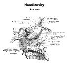
Nasal cavity


The nasal cavity forms part of the aerodigestive tract.
Gross anatomy
The nasal cavity is formed by :
- anteriorly: anterior nares
- laterally: inferior, middle and superior nasal conchae (turbinates)
- superiorly: cribriform plate of the ethmoid bone
- inferiorly: palatal processes of the maxilla and horizontal portion of the palatine bone forming the hard palate
- posteriorly: posterior nasal aperture (choanae) at the posterior margin of the bony nasal septum
In the midline, the nasal cavity is divided into right and left halves by the nasal septum composed of fibrocartilage anteriorly and the vomer and the perpendicular plate of the ethmoid bone posteriorly and inferiorly.
Anterior most, lies a small dilated portion, the nasal vestibule, which lies between the nasal aperture and the anterior nares, and posteriorly it is continuous with the nasopharynx via the posterior choanae. Laterally, the three nasal conchae form the three nasal meati.
Connections
- superior meatus
- posterior ethmoidal air cells and sphenoid sinuses via the sphenoethmoidal recess
- middle meatus
- frontal sinus via frontal recess
- anterior ethmoidal air cells and maxillary sinuses via ostiomeatal complex
- inferior meatus
Blood supply
Arterial supply
The arterial supply of the nasal cavity is rich and derives from both the internal and external carotid arteries:
- lateral nasal wall
- superior lateral nasal wall: anterior and posterior ethmoidal arteries
- inferior and middle turbinates: sphenopalatine artery
- posterior lateral nasal wall: pharyngeal artery
- nasal septum
- anteriorly: greater palatine artery
- posteroinferiorly: sphenopalatine artery
- superiorly: anterior and posterior ethmoidal arteries
- floor
- greater palatine and superior labial arteries
Rich arterial supply results in two anastomotic areas, which are common sites of epistaxis :
- Woodruff area: anastomosis of sphenopalatine and pharyngeal arteries in the inferior lateral nasal wall, posterior to the inferior turbinate
- Kiesselbach plexus: anastomosis of the anterior ethmoid, greater palatine, sphenopalatine and superior labial arteries in the anteroinferior nasal septum (see article on Kiesselbach plexus)
Venous drainage
A rich venous submucosal venous network is formed by veins that accompany arteries. It should be noted that the posterior ethmoid veins anastomose with veins of the dura mater and orbit, making this a potential route of spread of infection. There is also an anastomosis with veins of the external nose .
Innervation
The olfactory nerve (CN I) supplies the special sensation of smell to the olfactory epithelium in the roof of the nasal cavity, with fibers passing upwards through the cribriform plate to the olfactory bulb.
Mucosal somatic sensation of the nasal cavity is derived from numerous nerves, but in general terms the branches of the ophthalmic division of the trigeminal nerve (CN Va) supply the anterosuperior half whereas branches of the maxillary division of the trigeminal nerve (CN Vb) supply the posteroinferior half. More specifically:
- the nasal septum is innervated by:
- anterior: anterior ethmoidal nerve
- posterosuperior: medial branch of the posterior superior nasal nerve
- posteroinferior: nasopalatine nerve
- the lateral nasal wall is innervated by:
- anterosuperior: anterior ethmoidal nerve
- anteroinferior: anterior superior alveolar nerve
- posterosuperior: lateral branch of the posterior superior nasal nerve
- posteroinferior: lateral posterior inferior nasal nerve from the greater palatine nerve
Lymphatic drainage
- anterior drainage: to the external nose
- posterior drainage: via separate pathways to the lateral pharyngeal lymph nodes or deep cervical chain

