noduläre regenerative Hyperplasie

Fatal liver
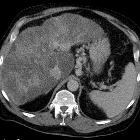
mass rupture in a common-variable-immunodeficiency patient with probable nodular regenerating hyperplasia. Hepatic imaging A–D. The initial multiphasic CT. A The non-contrasted image shows no abnormal intrahepatic bleeding. B The arterial phase shows multiple poorly-defined, arterial-enhancing nodules scattered throughout both hepatic lobes (white arrows). In portovenous phase C and 5 min-delayed phase D, these nodules remain iso- to hyperattenuating compared to the adjacent liver parenchyma. Hepatic imaging E–F. The 7-month follow-up CT. E The non-contrasted image shows multiple areas of intrahepatic bleeding (black arrowheads) and intraperitoneal bleeding (black arrows). F The portovenous phase demonstrates disruption of the liver capsule (white arrowhead), suggesting rupture of these hypervascular liver lesions
noduläre regenerative Hyperplasie
Siehe auch:
- hypervaskularisierte Leberläsionen
- Leberhämangiom
- Leberzirrhose
- Regeneratknoten der Leber
- hepatozelluläres Karzinom
- Leberadenom
- Fokale noduläre Hyperplasie
- fibrolamelläres hepatozelluläres Karzinom
- adenomatöse Hyperplasie Leber
- dysplastische Leberknoten
- multiple FNH syndrome
- nodular regenerative hyperplasia in chronic Budd-Chiari syndrome
- focal nodular hyperplasia vs. HCC
- hyper vascular hepatic metastasis
und weiter:

 Assoziationen und Differentialdiagnosen zu noduläre regenerative Hyperplasie:
 nicht verwechseln mit:


