Oppenheimers Ossikel

Oppenheimer
ossicle • Oppenheimer ossicle: illustrations - Ganzer Fall bei Radiopaedia

Oppenheimer
ossicle • Oppenheimer ossicle - Ganzer Fall bei Radiopaedia

Oppenheimer
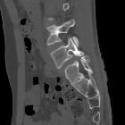
ossicle - a tricky rare variant!. CT sagittal view demonstrates a well-corticated bony fragment at the posterio-inferior aspect of L4-L5 facetal joint.

Oppenheimer
ossicle - a tricky rare variant!. CT sagittal view demonstrates sacralisation of L5. Also no cortical break or wedging is noted. No abnormal soft tissue component is seen.

Oppenheimer
ossicle - a tricky rare variant!. Well-corticated bony fragment along inferior articular facet on left side.

Oppenheimer
ossicle - a tricky rare variant!. 3D VR images clearly demonstrate the ossicle at the level of left inferior articular facet.

Oppenheimer
ossicle • Oppenheimer ossicle - Ganzer Fall bei Radiopaedia

Oppenheimer
ossicle • Oppenheimer's ossicle - Ganzer Fall bei Radiopaedia

Oppenheimer
ossicle • Oppenheimer ossicle - Ganzer Fall bei Radiopaedia

Oppenheimer
ossicle • Oppenheimer ossicle - Ganzer Fall bei Radiopaedia

Oppenheimer
ossicle • Oppenheimer ossicle - Ganzer Fall bei Radiopaedia
Oppenheimer ossicles are accessory ossicles associated with the facet joints found in ~4% (range 1-7%) of lumbar spines.
Oppenheimer ossicles are thought to arise as a result of non-union of a secondary ossification center of the articular process. They predominantly occur as a single, unilateral ossicle of the inferior articular processes of the lumbar spine although it can also occur at the superior articular process.
History and etymology
Oppenheimer nodules were first described by Albert Oppenheimer in 1942 .
Differential diagnosis
- fracture of the articular process: an Oppenheimer ossicle will demonstrate regular, well-defined corticated margins
Siehe auch:

 Assoziationen und Differentialdiagnosen zu Oppenheimers Ossikel:

