orbital dermoid cyst


Orbital dermoid cysts are congenital lesions representing closed sacs lined by an ectodermal epithelium and comprising the most common orbital mass in children. They are typically divided into deep (within the orbit) and superficial (adjacent to the orbital rim).
Epidemiology
They comprise ~2% of orbital tumors . Superficial location is much more frequent.
Clinical presentation
Superficial angular dermoid is usually diagnosed relatively early. As they grow slowly, less than 25% of them are identified at birth, and they usually manifest in the first decade of life. Clinical features include a painless subcutaneous mass along the zygomaticofrontal and the frontoethmoidal sutures .
It is important to note that more than 80% occur in the upper outer quadrant or the lacrimal fossa (external angular dermoid) .
Deep dermoids tend to be diagnosed later in life with accompanying proptosis .
Pathology
Dermoid cysts are thought to occur as a developmental anomaly in which embryonic ectoderm is trapped in the closing neural tube between the 5th-6th weeks of gestation .
Stratified squamous epithelium lines dermoid cysts, as in epidermoid cysts. Unlike epidermoid cysts, however, they also have epidermal appendages such as hair follicles, sweat glands, and sebaceous glands. The latter is responsible for the secretion of sebum which imparts the characteristic appearance of these lesions on CT and MRI.
A common misconception is that dermoid cysts contain adipose tissue. This is not the case, as lipocytes are mesodermal in origin, and dermoid cysts (by definition) are purely ectodermal. A dermoid cyst with adipose tissue would be a teratoma.
Radiographic features
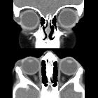
These lesions are usually extraconal, non-enhancing masses with smooth margins, cystic and/or solid components. They are typically heterogeneous with soft tissue, fluid and fatty (sebum) components; occasionally calcifications may be present.
They are most commonly located superotemporally, arising from the zygomaticofrontal suture, followed by superonasally, arising from the frontoethmoidal or frontolacrimal sutures.
Ruptured dermoids may show adjacent inflammatory changes.
Treatment and prognosis
Treatment and prognosis depend on size, location and involvement of orbital structures. While superficial lesion may barely require a cosmetic excision, a deeper one may require more invasive methods involving micro-dissection, orbitotomy, and rarely, intracranial exploration if the lesion extends to that extent .
Differential diagnosis
Possible differential considerations include
- orbital epidermoid cyst
- orbital teratoma
- usually large lesions and associated with a facial deformity
- multiloculated cystic masses
- calcification, fat, and/or ossification may be present
- orbital dermolipoma
- orbital hemangioma
See main article
Siehe auch:
und weiter:


 Assoziationen und Differentialdiagnosen zu Dermoidzyste der Orbita:
