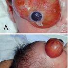
orbital teratoma

Orbitales,
retrobulbäres Teratom bei einem 2 Monate alten Kind, somit kongenital: Oben T2 axial, T1 axial, T2 koronar; unten T1 KM 3 Ebenen

Congenital
orbital teratoma: a case report with preservation of the globe and 18 years of follow-up. a and b The 1-day infant at presentation

Congenital
orbital teratoma: a case report with preservation of the globe and 18 years of follow-up. a CT of the orbits shows a large heterogeneous mass, filling the entire orbit. b T2-weighted MRI shows the tumor to be hyperintense to fat and extraocular muscles

Congenital
orbital teratoma in fetal MRI. Axial CT reveals enlargement of the left orbit containing a predominantly cystic mass. Minor associated calcification in the posterior globe. No identifiable lens or anterior chamber.
 Assoziationen und Differentialdiagnosen zu intraorbitales Teratom:
