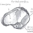
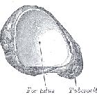
Os naviculare pedis


Os tibiale
externum - Klassifikation nach Geist: Von links nach rechts Typ 1 bis 3.

Os tibiale
externum im Röntgenbild des Fußes, hier Typ 1 nach Geist, also ein runder, vom Os naviculare isolierter Knochen.

Os tibiale
externum im Röntgenbild des Fußes, hier Typ 2 nach Geist, also ein dreieckiger, mit dem Os naviculare in breitem Kontakt stehender Knochen.

Dorsoplantar
X-ray of the feet of a 44 year old woman with suspected pes cavus. The right foot (to the left in the image) has an accessory navicular bone, type 2. On the left foot (to the right in the image) it is fused with the navicular bone, forming a cornuate navicular bone (white arrow), which is type 3.

Os naviculare
cornutum im Röntgenbild: Hakenförmige Ausziehung des medialen Rands des Os naviculare wie ein verschmolzenes Os tibiale externum (siehe Geist-Klassifikation).

Os naviculare
cornutum im Röntgenbild: Hakenförmige Ausziehung des medialen Rands des Os naviculare wie ein verschmolzenes Os tibiale externum (siehe Geist-Klassifikation).
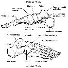
Talus •
Lower limb bones (illustrations) - Ganzer Fall bei Radiopaedia

Tarsal bones
• Navicular (Gray's illustration) - Ganzer Fall bei Radiopaedia


 Assoziationen und Differentialdiagnosen zu Os naviculare pedis:


