ossäre Lymphominfiltration

Lymphombefall
des Knochens (Femur links). Typische Lodwick 3-Läsion. Deutliche Periostreaktion.

Pathologische
Fraktur des Humerus bei ossärer Manifestation eines Non-Hodgkin-Lymphoms.

Lymphombefall
des Knochens (Femur links). Typische Lodwick 3-Läsion. Deutliche Periostreaktion.

Spectrum of
imaging findings in AIDS-related diffuse large B cell lymphoma. Coronal and axial CECT images of the abdomen (a and b) show hepatomegaly and a heterogenous hypoattenuating mass infiltrating through most of the right lobe of the liver. The infiltrative liver mass had more focal hypoattenuating areas (fat arrow). Aortocaval lymph nodes are also noted (short arrow). Axial CT through the pelvis in bone window (c) shows a permeative process involving the left femoral head and neck and associated pathological fracture

Spectrum of
imaging findings in AIDS-related diffuse large B cell lymphoma. CECT of the pelvis in soft tissue window (a) and bone window (b). A mixed lytic and sclerotic process involving the sacrum and extending to the iliac bones through the sacroiliac joints is seen. There are associated pathological fractures and a large extra osseous soft tissue component. There are also enlarged necrotic bilateral pelvic sidewall and inguinal lymph nodes

Primary
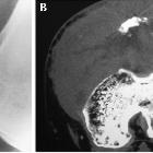
tumors of the patella. Patellar lymphoma with a pathologic fracture. (A) On radiographs, the patella consists of a sclerotic proximal portion and a lytic distal portion. (B) Permeative bone destruction, hazy margins, destroyed cortex, large soft tissue mass surrounding the patella, and joint involvement are shown on CT images.

Secondary
involvement of the bone with lymphoma • Non Hodgkin's lymphoma with metastasis to spine - Ganzer Fall bei Radiopaedia

Secondary
involvement of the bone with lymphoma • Secondary bone lymphoma - Ganzer Fall bei Radiopaedia

Secondary
involvement of the bone with lymphoma • Secondary bone lymphoma - Ganzer Fall bei Radiopaedia
Secondary involvement of the bone with lymphoma, also referred as secondary bone lymphoma, is much more common than primary bone lymphoma, occurring in ~15% of disseminated lymphomas.
Terminology
Secondary bone lymphoma is defined as lymphoma involving the bone with nodal disease occurring within six months or secondary lymphoma involving the bone at least six months after diagnosis.
Epidemiology
Secondary bone involvement is more common in children (25%) .
Clinical presentation
Bone pain and/or pathological fracture
Pathology
Secondary bone involvement can result from direct spread from nodal disease or haematogenous metastases. The axial skeleton is more often affected than the appendicular skeleton . The most frequent locations are :
- spine
- bony pelvis
- skull
- ribs
- facial bones
Radiographic features
Plain radiograph / CT
- lytic lesion(s)
- permeative bone destruction with cortical breach
- adjacent soft tissue mass(es)
Siehe auch:
- pathologische Fraktur
- Lymphom
- Ewing-Sarkom
- Non-Hodgkin-Lymphom
- diffuse skelettale Sklerosierung
- primäres Lymphom des Knochens
- NHL mit skelettaler Beteiligung
- Hodgkin-Lymphom des Knochens
und weiter:

 Assoziationen und Differentialdiagnosen zu Skeletaler Lymphombefall:


