Lymphom


Lymphoma (historically lymphosarcoma was used for diffuse forms of the disease) is a malignancy arising from lymphocytes or lymphoblasts. Lymphoma can be restricted to the lymphatic system or can arise as extranodal disease. This, along with variable aggressiveness results in a diverse imaging appearance.
Epidemiology
Lymphoma accounts for ~4% of all cancers . They are more common in developed countries.
In children, lymphoma accounts for 10-15% of all cancers, being the third most common form of malignancy .
Clinical presentation
Lymphoma can present as nodal or extranodal disease. Hodgkin lymphoma and low-grade non-Hodgkin lymphoma (NHL) classically present as nodal disease, whereas high-grade NHL can present with complications from mass effect such as superior vena cava obstruction, cauda equina syndrome, etc. Extranodal disease can affect any organ.
Lymphoma can often present with B symptoms (fever, night sweats and weight loss).
Pathology
Lymphomas are a malignancy that arise from mature lymphocytes. The etiology is unknown but potential lymphomatogenic risk factors include :
- viral infection, e.g. EBV, HTLV-1, HIV, HCV, HSV
- bacterial infection, e.g. Helicobacter pylori
- chronic immunosuppression, e.g. post-transplantation
- prior chemotherapy (especially alkalising agents) and drug therapy, e.g. digoxin
Classification
Lymphomas are currently classified according to the 2008 WHO classification of tumors of hematopoietic and lymphoid tissues. The main division is into:
- Hodgkin lymphoma (Hodgkin disease) (40%)
- non-Hodgkin lymphoma (60%)
- mature B-cell lymphoma
- mature T-cell and NK-cell lymphoma
- post-transplant lymphoproliferative disorders
The majority (85%) of lymphomas are B-cell with the remainder (15%) being T-cell .
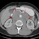
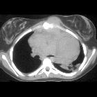
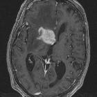
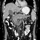
Location
Additionally, it is worth, especially for radiologists, dividing extranodal lymphomas according to the location:
- central nervous system (CNS)
- head and neck lymphoma
- thoracic lymphoma
- gastrointestinal lymphoma
- hepatobiliary lymphoma
- musculoskeletal lymphoma
- cutaneous lymphoma
- genitourinary lymphoma
- multi-regional
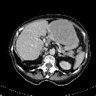
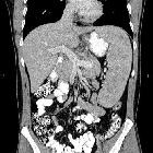
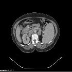
Radiographic features
Imaging characteristics will depend on the location and subtype of lymphoma. CT is the workhorse of imaging in lymphoma and plays a crucial role in staging (see main article: lymphoma staging). US and MRI are also used; for example, when assessing cervical lymph nodes (US) or CNS lymphoma (MRI). FDG-PET is used for staging and re-staging of lymphoma.
Treatment and prognosis
Lymphoma cure rates are comparatively high (up to 90%) compared to many other malignancies. Prognosis depends not only on histological subtype and grade but also on stage, hence why imaging plays a pivotal role in treatment. Aggressive lymphomas (e.g. Burkitt lymphoma) typically have a prognosis of weeks without treatment.
Siehe auch:
- orbitales Lymphom
- primäres ZNS-Lymphom
- mediastinal lymphoma
- Morbus Hodgkin
- Lymphom pulmonale Manifestation
- ZNS Lymphom
- Lymphombefall des Pankreas
- Lymphom des Magens
- sekundäres ZNS-Lymphom
- Seminom Metastasierung
- vaginal lymphoma
- Lymphom des spinalen Myelons
- primäres Dünndarmlymphom
- Lymphom der Zervix uteri
- Lymphom des Uterus
- WHO classification of neoplastic diseases of the lymphoid tissues
und weiter:
- Osteomyelitis
- Lungenkarzinom
- verkalkte mediastinale Lymphknoten
- T2 hyperintense Basalganglien
- carotid space
- Tumoren des vorderen oberen Mediastinums
- zystische Lymphknoten
- Tumor Kleinhirnbrückenwinkel
- Lymphom der Niere
- solitärer pulmonaler Rundherd
- Chylothorax
- Morbus Castleman
- Tumoren des hinteren Mediastinums
- Vergrößerung der zervikalen Lymphknoten
- Thymuslipom
- Interstitielle Lungenerkrankung
- posterior mediastinal masses
- zerebrale Läsionen mit ringförmiger Kontrastmittelanreicherung
- primary hepatic lymphoma
- differential of chronic alveolar opacities
- bilaterale axilläre Lymphadenopathie
- choline:creatine ratio
- adenocarcinoma of the small bowel
- duodenal filling defects
- anterosuperior mediastinal mass (mnemonic)
- Lymphom der Mamma
- Skrofeln
- Krebs
- Burkitt-Lymphom
- chronic bilateral airspace opacification
- differential diagnosis of unilateral axillary lymphadenopathy
- musculoskeletal manifestations of AIDS
- Raumforderungen im oder am Sinus cavernosus
- differential of a large unilateral pleural effusion
- Lymphom der Schilddrüse
- acute airspace opacification with lymphadenopathy
- Knochenläsionen der Diaphyse
- peritoneal lymphomatosis secondary to gastric lymphoma
- Lymphom Kaverne
- eosinophilic hyperplastic lymphogranuloma
- vertebral involvement in Burkitt's lymphoma
- lymphoma of the urinary system
- mediastinal T-cell lymphoblastic lymphoma
- primary bronchial lymphoma
- HIV-related primary hepatic lymphoma with renal involvement
- Lymphom am Fuß
- Lymphom der Dura
- Lymphombefall Extremitäten
- Ursachen für Perfusionsdefekte in der Lungenventilations / -perfusionsszintigraphie
- Meningeosis lymphomatosa
- chronische Periaortitis


 Assoziationen und Differentialdiagnosen zu Lymphom: