carotid space


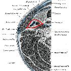
The carotid space, the suprahyoid portion of which is also known as the poststyloid parapharyngeal space, is a deep compartment of the head and neck bound by the carotid sheath.
Terminology
The "carotid space" terminology was introduced by some radiologists to facilitate differential diagnosis and is used in this article . However, in much of the surgical and anatomical literature, the carotid space within the suprahyoid neck is considered part of the parapharyngeal space . Under this nomenclature, the parapharyngeal space is divided into prestyloid and poststyloid (retrostyloid) compartments by the tensor-vascular-styloid fascia . The poststyloid parapharyngeal space consists of or includes the carotid space or sheath around the cervical internal carotid artery.
Gross anatomy
The carotid space is a roughly cylindrical space that extends from the skull base through to the aortic arch. It is circumscribed by the carotid sheath, which has contributions from all three layers of the deep cervical fascia . Above the carotid bifurcation, the contribution of the middle layer of cervical fascia can be inconsistent , and the sheath is interrupted .
The carotid artery bifurcation occurs near the level of the hyoid bone .
Contents
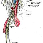
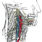
- common carotid artery inferiorly and internal carotid artery superiorly
- internal jugular vein
- ansa cervicalis (embedded in the anterior wall of the sheath)
- nerves
- vagus nerve (CN X): posterior to vessels in posterior notch; extends below hyoid to mediastinum within the carotid sheath
- in the upper part of the carotid sheath, there is also
- glossopharyngeal nerve (CN IX): anterior to vessel
- accessory nerve (CN XI)
- hypoglossal nerve (CN XII)
- sympathetic nerves: medial to vessels and lateral to the retropharyngeal space
- deep cervical lymph node chain (levels II, III, and IV)
Relations
Suprahyoid carotid space:
- anteriorly: masticator space; parapharyngeal space
- laterally: parotid space
- posteriorly: perivertebral space
- medially: retropharyngeal space
The suprahyoid portion of the carotid space is synonymous with the poststyloid compartment of the parapharyngeal space.
Infrahyoid carotid space:
- anteriorly: anterior cervical space
- laterally: posterior cervical space
- posteriorly: perivertebral space
- medially: retropharyngeal and visceral space
Boundaries
- superior margin: lower border of jugular foramen
- inferior margin: aortic arch
The carotid sheath is made from the various regional fascia, including contributions from all three layers of the deep cervical fascia :
- lateral margin: fascia of the sternocleidomastoid muscle (superficial layer of the deep cervical fascia)
- anterior margin: stylopharyngeal aponeurosis or tensor-vascular-styloid fascia (middle layer of the deep cervical fascia)
- medial margin: cloison sagittale (either part of the deep or middle layer of the deep cervical fascia)
- posterior margin: prevertebral fascia and/or alar fascia (deep layer of the deep cervical fascia)
Radiographic appearance
Ultrasound
Ultrasound is good for identifying vascular structures in the lateral neck. The origin of the internal and external carotid arteries is the main point of sonographic interest as the carotid space superior to this cannot be visualized due to the mastoid process.
The internal carotid is usually posterolateral to the external carotid however in 5-10% of the population the internal carotid lies medial or anterior or the two vessels may lie within the same frontal plane .
The external carotid can be identified from the internal carotid as:
- it branches within the neck; the first immediate branch is the superior thyroid artery
- Doppler tracing of vessel flow within the internal carotid is typical of low peripheral resistance (high diastolic peak), whereas the external carotid has high-resistance flow (lower diastolic peak, systolic peak higher than that of the internal carotid)
CT
- often modality of choice when evaluating for pathology within the carotid space because CT is able to identify any benign or malignant tumors within the carotid space and allows for better visualization of any associated bone involvement in comparison to MRI
- best identifies vascular lesions on CT angiography
MRI
- useful for identifying between the different types of benign tumors involving the carotid space
- provides better soft tissue contrast in comparison to CT and can be useful at elucidating any signs of intramural metastatic nodal invasion of the carotid artery which is a relative contraindication for surgical tumor resection
Related pathology
A mass originating from the carotid space will cause anterior displacement of the parapharyngeal space fat. Lesions include :
- tumors
- neurogenic tumors (most common): paraganglioma, schwannoma, neurofibroma
- lymph nodes: metastatic lymphadenopathy, lymphoma
- meningioma
- vascular lesions
- cellulitis/abscess
Siehe auch:
- Arteria carotis interna
- Meningeom
- Arteria carotis communis
- Lymphom
- Schwannom
- Paragangliom
- Neurofibrom
- parotid space
- deep compartments of the head and neck
- Nervus glossopharyngeus
- masticator space
- Retropharyngealraum
- Nervus hypoglossus
- superficial mucosal space
- Nervus accessorius
- prevertebral space
- Halsfaszie
- Parapharyngealraum
und weiter:

 Assoziationen und Differentialdiagnosen zu carotid space:


