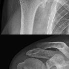
osteonecrosis of the humeral head

Osteonecrosis
• Osteonecrosis of humeral and femoral heads - Ganzer Fall bei Radiopaedia

Osteonecrosis
• Avascular necrosis - Ganzer Fall bei Radiopaedia

Dysbaric
osteonecrosis of the humerus. Coronal T1-WI (A and B): intramedullary lesion with geographical borders within the proximal diaphysis (red arrow), with epiphyseal extension (black arrow).

Humeral head
osteonecrosis in an adolescent amateur swimming athlete: a case report. An anteroposterior plain radiograph (2007-5-25) shows the lesion became larger and a sclerotic band appeared around the lesion.

Humeral head
osteonecrosis in an adolescent amateur swimming athlete: a case report. A coronal oblique T1-weighted magnetic resonance image of the shoulder (2007-5-25) (spin-echo sequence with a repetition time of 626 msec and an echo time of 13 msec) shows a focal low signal area in the medial part of humeral head (A), and a coronal oblique T2-weighted magnetic resonance image of the shoulder (2007-5-25) (spin-echo sequence with a repetition time of 3775 msec and an echo time of 90 msec) shows an irregular low signal band on the corresponding part of humeral head (B).

Avascular
necrosis of humeral head in an elderly patient with tuberculosis: a case report. Computed tomography scan at the level of the upper humerus showing crescentic lucency as evidence of osteonecrosis.

Osteonecrosis
of the humeral head in a human immunodeficiency virus-infected patient under tenofovir disoproxil fumarate–emtricitabine–lopinavir/ritonavir for 10 years: a case report. The left shoulder x-ray showed a patch of osteolysis (thick blue arrow) on the humeral head with a clear osteosclerosis border line (small black arrow). The lesion is centered by an osteocondensed image (long black arrow) with an appearance of cortical rupture typical of systemic osteonecrosis (red arrow)

A case of
completed course multifocal osteonecrosis (MFON) during pregnancy due to primary antiphospholipid syndrome. MRI of both shoulders revealed abnormal signals in the right humeral head involving the superior-posterior aspect which are well demarcated from the adjacent normal bony by a thin rim of low signal material in both T1 and T2 WI. No evidence of structural collapse with marrow edema showing high signal with fat sat sequence. Conclusion: grade II AVN of the right humeral head and grade I AVN of the left humeral head with minimal joint effusion

Osteonecrosis
• Post-traumatic osteonecrosis of the humeral head - Ganzer Fall bei Radiopaedia

Osteonecrosis
• Humeral head and femoral head osteonecrosis due to sickle cell disease - Ganzer Fall bei Radiopaedia

Osteonecrosis
• Avascular necrosis of the humeral head - Ganzer Fall bei Radiopaedia

Radiography
of total avascular necrosis of right humeral head. Woman of 81 years old with diabetes of long evolution.

Scapular
fracture • Bilateral humeral head avascular necrosis - Ganzer Fall bei Radiopaedia

Osteonecrosis
• Avascular necrosis of humerus - Ganzer Fall bei Radiopaedia

Osteonecrosis
• Post-traumatic avascular necrosis of the humeral head - Ganzer Fall bei Radiopaedia

Osteonecrosis
of the humeral head • Osteonecrosis of the humeral head - Ganzer Fall bei Radiopaedia

Osteonecrosis
of the humeral head • Osteonecrosis of the humeral head - Ganzer Fall bei Radiopaedia

Osteonecrosis
of the humeral head • Avascular necrosis of the shoulder - Cruess stage I - Ganzer Fall bei Radiopaedia

Osteonecrosis
of the humeral head • Osteonecrosis of the humeral head - Ganzer Fall bei Radiopaedia

Dysbaric
osteonecrosis of the humerus. Anteroposterior (AP) radiograph of the right shoulder with the humerus in external rotation shows no abnormalities.

Dysbaric
osteonecrosis of the humerus. MR arthrography, coronal fs T1-WI (A and B): irregularity in the supraspinatus (white arrow), bone marrow abnormalities in the humerus, with low SI centrally (white asterisk) and intermediate to high SI peripherally (red arrow).

Dysbaric
osteonecrosis of the humerus. MR arthrography, coronal fs T2-WI (A and B): the peripheral borders are strongly hyperintense (red arrow), while the center of the lesion remains hypointense (white asterisk).

Dysbaric
osteonecrosis of the humerus. MR arthrography, coronal T2-WI: double-line sign with inner hyperintense rim (black arrow) and outer hypointense rim (red arrow).

Osteonecrosis
of the humeral head • Post-traumatic osteonecrosis of the proximal humerus after Salter Harris I fracture - Ganzer Fall bei Radiopaedia
Osteonecrosis of the humeral head (ONHH), also known as Hass disease, is considered the second commonest location for osteonecrosis (following the hip).
Pathology
It generally develops in the subchondral region. In some patients, osteonecrosis can lead to collapse of the necrotic subchondral bone, development of an irregular joint surface, and subsequent joint degeneration.
The general risk factors for osteonecrosis, in general, apply to humeral head osteonecrosis. Corticosteroid use has been recognized as a dominant causative association .
Radiographic features
While many of the general radiographic features for osteonecrosis may be present with ONHH, the presence of crescent sign can be the classic diagnostic feature in the correct clinical context .
See also
Siehe auch:
- Aseptische Knochennekrose
- crescent sign
- Cruess-Klassifikation der aseptischen Nekrose des Humeruskopfes
- Glenohumeral osteoarthritis
und weiter:

 Assoziationen und Differentialdiagnosen zu Aseptische Knochennekrose des Humeruskopfes:

